

Proteomics study of human cord blood reticulocyte-derived exosomes
- PMID: 30232403
- PMCID: PMC6145868
- DOI: 10.1038/s41598-018-32386-2
Proteomics study of human cord blood reticulocyte-derived exosomes
Abstract
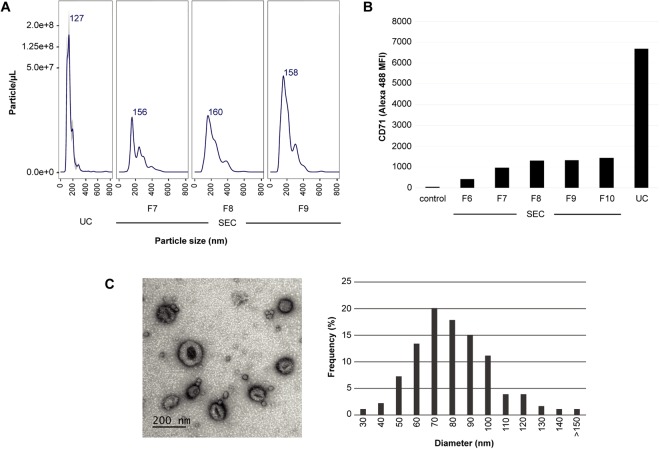
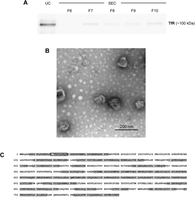
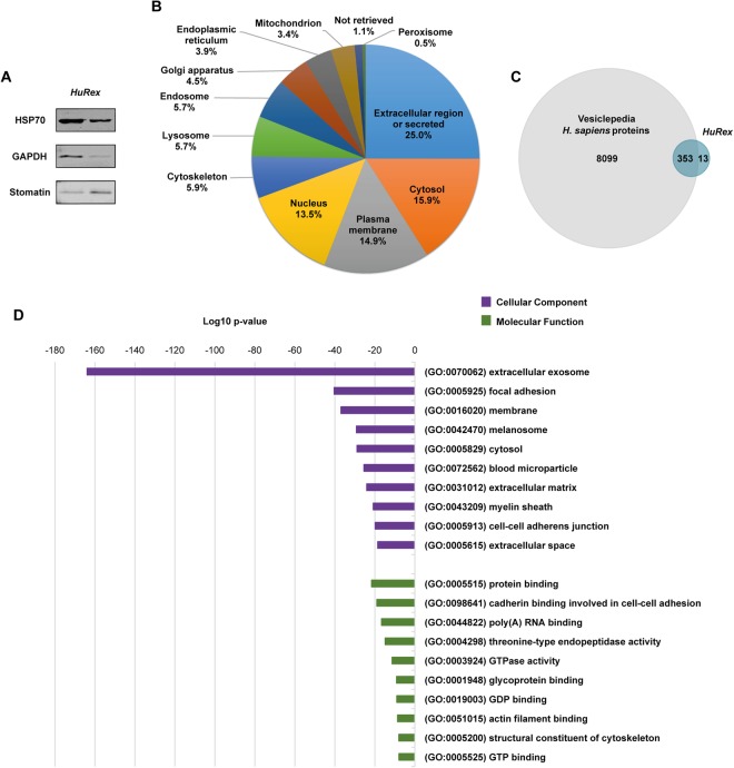
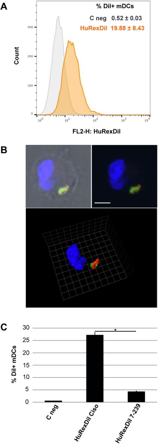
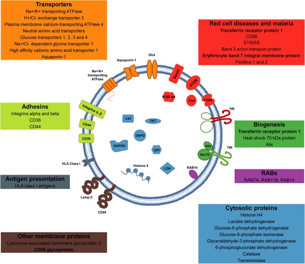
Reticulocyte-derived exosomes (Rex), extracellular vesicles of endocytic origin, were initially discovered as a cargo-disposal mechanism of obsolete proteins in the maturation of reticulocytes into erythrocytes. In this work, we present the first mass spectrometry-based proteomics of human Rex (HuRex). HuRex were isolated from cultures of human reticulocyte-enriched cord blood using different culture conditions and exosome isolation methods. The newly described proteome consists of 367 proteins, most of them related to exosomes as revealed by gene ontology over-representation analysis and include multiple transporters as well as proteins involved in exosome biogenesis and erythrocytic disorders. Immunoelectron microscopy validated the presence of the transferrin receptor. Moreover, functional assays demonstrated active capture of HuRex by mature dendritic cells. As only seven proteins have been previously associated with HuRex, this resource will facilitate studies on the role of human reticulocyte-derived exosomes in normal and pathological conditions affecting erythropoiesis.
Conflict of interest statement
The authors declare no competing interests.
Figures
Similar articles
-
Quantitative mass spectrometry of human reticulocytes reveal proteome-wide modifications during maturation.Br J Haematol. 2018 Jan;180(1):118-133. doi: 10.1111/bjh.14976. Epub 2017 Nov 2. Br J Haematol. 2018. PMID: 29094334
-
Comparison of the Proteome of Adult and Cord Erythroid Cells, and Changes in the Proteome Following Reticulocyte Maturation.Mol Cell Proteomics. 2016 Jun;15(6):1938-46. doi: 10.1074/mcp.M115.057315. Epub 2016 Mar 22. Mol Cell Proteomics. 2016. PMID: 27006477 Free PMC article.
-
Extrusion of Na,K-ATPase and transferrin receptor with lipid raft-associated proteins in different populations of exosomes during reticulocyte maturation in dogs.Jpn J Vet Res. 2010 May;58(1):17-27. Jpn J Vet Res. 2010. PMID: 20645582
-
The Jeanne Manery-Fisher Memorial Lecture 1991. Maturation of reticulocytes: formation of exosomes as a mechanism for shedding membrane proteins.Biochem Cell Biol. 1992 Mar-Apr;70(3-4):179-90. doi: 10.1139/o92-028. Biochem Cell Biol. 1992. PMID: 1515120 Review.
-
Inside(sight) of tiny communicator: exosome biogenesis, secretion, and uptake.Mol Cell Biochem. 2020 Apr;467(1-2):77-94. doi: 10.1007/s11010-020-03703-z. Epub 2020 Feb 22. Mol Cell Biochem. 2020. PMID: 32088833 Review.
Cited by
-
Cellular dynamics of mammalian red blood cell production in the erythroblastic island niche.Biophys Rev. 2019 Dec;11(6):873-894. doi: 10.1007/s12551-019-00579-2. Epub 2019 Aug 15. Biophys Rev. 2019. PMID: 31418139 Free PMC article. Review.
-
CD34+ HSPCs-derived exosomes contain dynamic cargo and promote their migration through functional binding with the homing receptor E-selectin.Front Cell Dev Biol. 2023 Apr 25;11:1149912. doi: 10.3389/fcell.2023.1149912. eCollection 2023. Front Cell Dev Biol. 2023. PMID: 37181754 Free PMC article.
-
Characterization and Proteomic Analysis of Plasma EVs Recovered from Healthy and Diseased Dogs with Canine Leishmaniosis.Int J Mol Sci. 2023 Mar 13;24(6):5490. doi: 10.3390/ijms24065490. Int J Mol Sci. 2023. PMID: 36982564 Free PMC article.
-
Toxoplasma gondii exploits the host ESCRT machinery for parasite uptake of host cytosolic proteins.PLoS Pathog. 2021 Dec 13;17(12):e1010138. doi: 10.1371/journal.ppat.1010138. eCollection 2021 Dec. PLoS Pathog. 2021. PMID: 34898650 Free PMC article.
-
Extracellular Vesicle-Based Therapeutics: Preclinical and Clinical Investigations.Pharmaceutics. 2020 Dec 1;12(12):1171. doi: 10.3390/pharmaceutics12121171. Pharmaceutics. 2020. PMID: 33271883 Free PMC article. Review.
References
Publication types
MeSH terms
Substances
LinkOut - more resources
Full Text Sources
Other Literature Sources
Molecular Biology Databases
Research Materials
