Protective effects of Brazilian propolis supplementation on capillary regression in the soleus muscle of hindlimb-unloaded rats
- PMID: 30232713
- PMCID: PMC10717714
- DOI: 10.1007/s12576-018-0639-z
Protective effects of Brazilian propolis supplementation on capillary regression in the soleus muscle of hindlimb-unloaded rats
Abstract
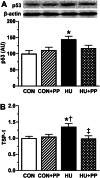
The protective effects of Brazilian propolis on capillary regression induced by chronically neuromuscular inactivity were investigated in rat soleus muscle. Four groups of male Wistar rat were used in this study; control (CON), control plus Brazilian propolis supplementation (CON + PP), 2-week hindlimb unloading (HU), and 2-week hindlimb unloading plus Brazilian propolis supplementation (HU + PP). The rats in the CON + PP and HU + PP groups received two oral doses of 500 mg/kg Brazilian propolis daily (total daily dose 1000 mg/kg) for 2 weeks. Unloading resulted in a decrease in capillary number, luminal diameter, and capillary volume, and an increase in the expression of anti-angiogenic factors, such as p53 and TSP-1, within the soleus muscle. Brazilian propolis supplementation, however, prevented these changes in capillary structure due to unloading through the stimulation of pro-angiogenic factors and suppression of anti-angiogenic factors. These results suggest that Brazilian propolis is a potential non-drug therapeutic agent against capillary regression induced by chronic unloading.
Keywords: Anti-angiogenic factors; Brazilian propolis; Capillary regression; Hindlimb unloading; Muscle atrophy; Oxidative stress.
Conflict of interest statement
The authors declare that they have no conflict of interest.
Figures


Similar articles
-
Preventive effects of nucleoprotein supplementation combined with intermittent loading on capillary regression induced by hindlimb unloading in rat soleus muscle.Physiol Rep. 2017 Feb;5(4):e13134. doi: 10.14814/phy2.13134. Epub 2017 Feb 27. Physiol Rep. 2017. PMID: 28242821 Free PMC article.
-
Amelioration of capillary regression and atrophy of the soleus muscle in hindlimb-unloaded rats by astaxanthin supplementation and intermittent loading.Exp Physiol. 2014 Aug;99(8):1065-77. doi: 10.1113/expphysiol.2014.079988. Epub 2014 Jun 6. Exp Physiol. 2014. PMID: 24907028
-
Protective effects of astaxanthin on capillary regression in atrophied soleus muscle of rats.Acta Physiol (Oxf). 2013 Feb;207(2):405-15. doi: 10.1111/apha.12018. Epub 2012 Oct 22. Acta Physiol (Oxf). 2013. PMID: 23088455
-
Protective effects of chlorogenic acid on capillary regression caused by disuse muscle atrophy.Biomed Res. 2021;42(6):257-264. doi: 10.2220/biomedres.42.257. Biomed Res. 2021. PMID: 34937825
-
Niacin supplementation attenuates the regression of three-dimensional capillary architecture in unloaded female rat skeletal muscle.Physiol Rep. 2024 Apr;12(8):e16019. doi: 10.14814/phy2.16019. Physiol Rep. 2024. PMID: 38627220 Free PMC article.
Cited by
-
Methylglyoxal reduces resistance exercise-induced protein synthesis and anabolic signaling in rat tibialis anterior muscle.J Muscle Res Cell Motil. 2024 Dec;45(4):263-273. doi: 10.1007/s10974-024-09680-w. Epub 2024 Jul 31. J Muscle Res Cell Motil. 2024. PMID: 39085712
-
Application of transcutaneous carbon dioxide improves capillary regression of skeletal muscle in hyperglycemia.J Physiol Sci. 2019 Mar;69(2):317-326. doi: 10.1007/s12576-018-0648-y. Epub 2018 Nov 26. J Physiol Sci. 2019. PMID: 30478742 Free PMC article.
-
Effects of astaxanthin supplementation and electrical stimulation on muscle atrophy and decreased oxidative capacity in soleus muscle during hindlimb unloading in rats.J Physiol Sci. 2019 Sep;69(5):757-767. doi: 10.1007/s12576-019-00692-7. Epub 2019 Jul 4. J Physiol Sci. 2019. PMID: 31273678 Free PMC article.
-
Effects of Propolis Extract and Propolis-Derived Compounds on Obesity and Diabetes: Knowledge from Cellular and Animal Models.Molecules. 2019 Dec 1;24(23):4394. doi: 10.3390/molecules24234394. Molecules. 2019. PMID: 31805752 Free PMC article. Review.
-
Green propolis extract attenuates acute kidney injury and lung injury in a rat model of sepsis.Sci Rep. 2021 Mar 15;11(1):5925. doi: 10.1038/s41598-021-85124-6. Sci Rep. 2021. PMID: 33723330 Free PMC article.
References
-
- de Leon EB, Bortoluzzi A, Rucatti A, Nunes RB, Saur L, Rodrigues M, Oliveira U, Alves-Wagner AB, Xavier LL, Machado UF, Schaan BD, Dall’Ago P. Neuromuscular electrical stimulation improves GLUT-4 and morphological characteristics of skeletal muscle in rats with heart failure. Acta Physiol (Oxf) 2011;201:265–273. doi: 10.1111/j.1748-1716.2010.02176.x. - DOI - PubMed
-
- Kanazashi M, Tanaka M, Murakami S, Kondo H, Nagatomo F, Ishihara A, Roy RR, Fujino H. Amelioration of capillary regression and atrophy of the soleus muscle in hindlimb-unloaded rats by astaxanthin supplementation and intermittent loading. Exp Physiol. 2014;99:1065–1077. doi: 10.1113/expphysiol.2014.079988. - DOI - PubMed
MeSH terms
Substances
Grants and funding
- No. 16H07361/Grants-in-Aid for Scientific Research from Japanese Ministry of Education, Culture, Sports, science and Technology
- No. 16K12732/Grants-in-Aid for Scientific Research from Japanese Ministry of Education, Culture, Sports, science and Technology
- No. 15K16516/Grants-in-Aid for Scientific Research from Japanese Ministry of Education, Culture, Sports, science and Technology
- N/A/Yamada Research Grant
LinkOut - more resources
Full Text Sources
Other Literature Sources
Medical
Research Materials
Miscellaneous

