. 2018 Nov;13(11):1974-1980.
doi: 10.4103/1673-5374.239445.

Hyperoside protects the blood-brain barrier from neurotoxicity of amyloid beta 1-42

Affiliations

Hyperoside protects the blood-brain barrier from neurotoxicity of amyloid beta 1-42

Chen-Yang Liu et al. Neural Regen Res. 2018 Nov.

Abstract

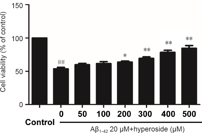
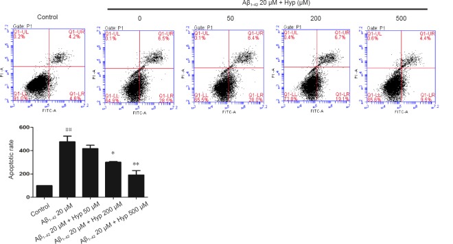
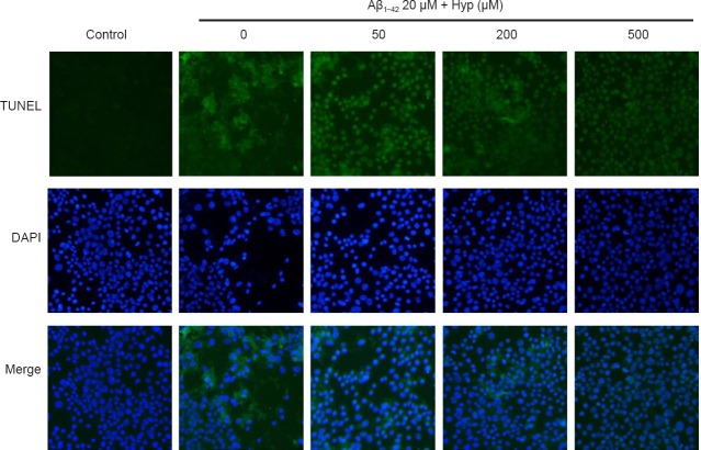
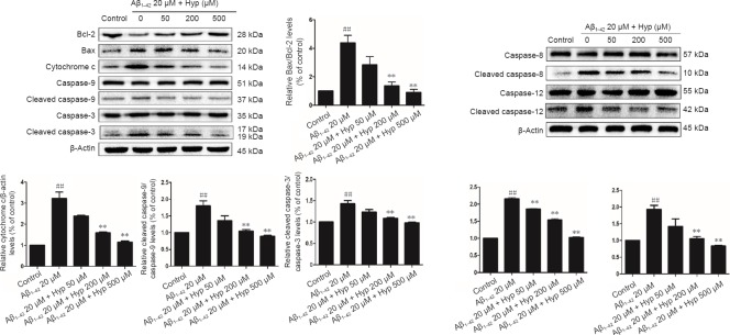
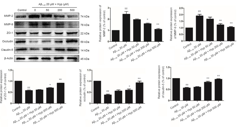
Mounting evidence indicates that amyloid β protein (Aβ) exerts neurotoxicity by disrupting the blood-brain barrier (BBB) in Alzheimer's disease. Hyperoside has neuroprotective effects both in vitro and in vivo against Aβ. Our previous study found that hyperoside suppressed Aβ1-42-induced leakage of the BBB, however, the mechanism remains unclear. In this study, bEnd.3 cells were pretreated with 50, 200, or 500 µM hyperoside for 2 hours, and then exposed to Aβ1-42 for 24 hours. Cell viability was determined using 3-(4,5-dimethyl-2-thiazolyl)-2,5-diphenyl-2-H-tetrazolium bromide assay. Flow cytometry and terminal deoxynucleotidyl transferase-mediated dUTP nick-end labeling assay were used to analyze cell apoptosis. Western blot assay was carried out to analyze expression levels of Bax, Bcl-2, cytochrome c, caspase-3, caspse-8, caspase-9, caspase-12, occludin, claudin-5, zonula occludens-1, matrix metalloproteinase-2 (MMP-2), and MMP-9. Exposure to Aβ1-42 alone remarkably induced bEnd.3 cell apoptosis; increased ratios of cleaved caspase-9/caspase-9, Bax/Bcl-2, cleaved caspase-8/caspase-8, and cleaved caspase-12/caspase-12; increased expression of cytochrome c and activity of caspase-3; diminished levels of zonula occludens-1, claudin-5, and occludin; and increased levels of MMP-2 and MMP-9. However, hyperoside pretreatment reversed these changes in a dose-dependent manner. Our findings confirm that hyperoside alleviates fibrillar Aβ1-42-induced BBB disruption, thus offering a feasible therapeutic application in Alzheimer's disease.

Keywords: Alzheimer's disease; amyloid beta 1-42; anti-apoptosis; bEnd.3 cells; blood-brain barrier; hyperoside; nerve regeneration; neural regeneration; tight junction proteins.


Conflict of interest statement

None

Figures

Figure 1
Figure 1
Effect of various concentrations of hyperoside on inhibiting Aβ1–42-induced damage in bEnd.3 cells. 3-(4,5-Dimethyl-2-thiazolyl)-2,5-diphenyl-2-H-tetrazolium bromide assay was used to detect the viability of bEnd.3 cells. Results were measured as optical density at 490 nm and presented as the mean ± SEM, and analyzed by one-way analysis of variance followed by Tukey's multiple comparison post hoc test. The experiment was conducted in triplicate. ##P < 0.01, vs. control group; *P < 0.05, **P < 0.01, vs. 20 µM Aβ1–42 group. Aβ1–42: Amyloid beta 1–42.
Figure 2
Figure 2
Protection of Hyp against Aβ1–42-induced apoptosis in bEnd.3 cells as detected by flow cytometry (FITC-V/PI) analysis. The apoptosis rate of fibrillar Aβ1–42-treated bEnd.3 cells obviously increased, and dramatically decreased after pretreatment with concentrations of Hyp from 50 to 500 µM. The results are presented as the mean ± SEM, and analyzed by one-way analysis of variance followed by Tukey's multiple comparison post hoc test. The experiment was conducted in triplicate. ##P < 0.01, vs. control group; *P < 0.05, **P < 0.01, vs. 20 µM Aβ1–42 group. Aβ1–42: Amyloid beta 1–42; Hyp: hyperoside.
Figure 3
Figure 3
Effect of Hyp on apoptotic cells in fibrillar Aβ1–42-induced bEnd.3 cells (fluorescence microscopy, 200× magnification). Apoptotic nuclei stained by TUNEL present green fluorescence and even nuclear fragmentation (red arrows). All nuclei are stained by DAPI with dark blue fluorescence. The experiment was conducted in triplicate. Aβ1–42: Amyloid beta 1–42; Hyp: hyperoside; TUNEL: terminal deoxynucleotidyl transferase-mediated dUTP nick-end labeling; DAPI: 4′,6-diamidino-2-phenylindole.
Figure 4
Figure 4
Hyp prevents Aβ1–42-induced apoptosis in bEnd.3 cells via inhibiting mitochondrial, endoplasmic reticulum, and death receptor signaling pathways. Results of western blot assay showed that ratios of Bax/Bcl-2, cleaved caspase-9/caspase-9, cleaved caspase-12/caspase-12, cleaved caspase-8/caspase-8, and cleaved caspase-3/caspase-3 were increased, while cytochrome c protein was reduced in Aβ1–42-induced apoptosis in bEnd.3 cells. Equal amount of protein loading was confirmed by actin levels. All results are represented as the mean ± SEM (one-way analysis of variance followed by Tukey's multiple comparison post hoc test). The experiment was conducted in triplicate. ##P < 0.01, vs. control group; **P < 0.01, vs. 20 µM Aβ1–42 group. Aβ1–42: Amyloid beta 1–42; Hyp: hyperoside.
Figure 5
Figure 5
Effect of Hypeon expression of TJs and MMPs in fibrillar Aβ1–42-treated bEnd.3 cells. Results of western blot assay showed expression of MMP-2, MMP-9 and TJs (ZO-1, occludin and claudin-5). Equal amount of protein loading was confirmed by β-actin levels. Data are represented as mean ± SEM (one-way analysis of variance followed by Tukey's multiple comparison post hoc test). The experiment was conducted in triplicate. ##P < 0.01, vs. control group; *P < 0.05, **P < 0.01, vs. 20 µM Aβ1–42 group. Aβ1–42: Amyloid beta 1–42; Hyp: hyperoside; TJs: tight junction proteins; MMP: matrix metalloproteinase; ZO-1: zonula occludens-1.

Similar articles

Cited by

References

    1. Abbott NJ, Patabendige AA, Dolman DE, Yusof SR, Begley DJ. Structure and function of the blood-brain barrier. Neurobiol Dis. 2010;37:13–25. - PubMed
    1. Bednarczyk J, Lukasiuk K. Tight junctions in neurological diseases. Acta Neurobiol Exp (Wars) 2011;71:393–408. - PubMed
    1. Biron KE, Dickstein DL, Gopaul R, Jefferies WA. Amyloid triggers extensive cerebral angiogenesis causing blood brain barrier permeability and hypervascularity in Alzheimer's disease. PLoS One. 2011;6:e23789. - PMC - PubMed
    1. Carrano A, Hoozemans JJ, van der Vies SM, Rozemuller AJ, van Horssen J, de Vries HE. Amyloid Beta induces oxidative stress-mediated blood-brain barrier changes in capillary amyloid angiopathy. Antioxid Redox Signal. 2011;15:1167–1178. - PubMed
    1. Chen X, Threlkeld SW, Cummings EE, Juan I, Makeyev O, Besio WG, Gaitanis J, Banks WA, Sadowska GB, Stonestreet BS. Ischemia-reperfusion impairs blood-brain barrier function and alters tight junction protein expression in the ovine fetus. Neuroscience. 2012;226:89–100. - PMC - PubMed