Review
. 2018 Aug 31:12:590.
doi: 10.3389/fnins.2018.00590. eCollection 2018.

In vitro Models for Seizure-Liability Testing Using Induced Pluripotent Stem Cells

Affiliations
Review

In vitro Models for Seizure-Liability Testing Using Induced Pluripotent Stem Cells

Alastair I Grainger et al. Front Neurosci. .

Abstract

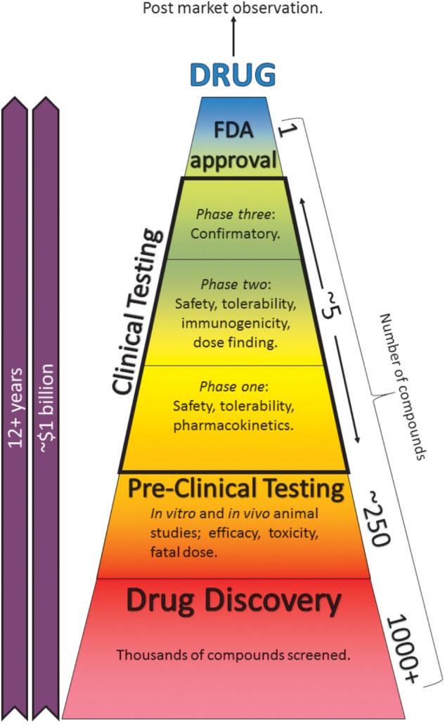
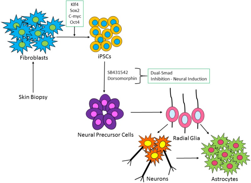
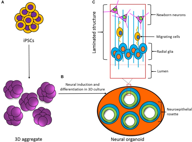
The brain is the most complex organ in the body, controlling our highest functions, as well as regulating myriad processes which incorporate the entire physiological system. The effects of prospective therapeutic entities on the brain and central nervous system (CNS) may potentially cause significant injury, hence, CNS toxicity testing forms part of the "core battery" of safety pharmacology studies. Drug-induced seizure is a major reason for compound attrition during drug development. Currently, the rat ex vivo hippocampal slice assay is the standard option for seizure-liability studies, followed by primary rodent cultures. These models can respond to diverse agents and predict seizure outcome, yet controversy over the relevance, efficacy, and cost of these animal-based methods has led to interest in the development of human-derived models. Existing platforms often utilize rodents, and so lack human receptors and other drug targets, which may produce misleading data, with difficulties in inter-species extrapolation. Current electrophysiological approaches are typically used in a low-throughput capacity and network function may be overlooked. Human-derived induced pluripotent stem cells (iPSCs) are a promising avenue for neurotoxicity testing, increasingly utilized in drug screening and disease modeling. Furthermore, the combination of iPSC-derived models with functional techniques such as multi-electrode array (MEA) analysis can provide information on neuronal network function, with increased sensitivity to neurotoxic effects which disrupt different pathways. The use of an in vitro human iPSC-derived neural model for neurotoxicity studies, combined with high-throughput techniques such as MEA recordings, could be a suitable addition to existing pre-clinical seizure-liability testing strategies.

Keywords: astrocytes; iPSC neurons; in vitro; safety pharmacology; seizures.


Figures

FIGURE 1
FIGURE 1
The drug-development process. Taking anywhere up to 15 years and a cost of ∼$1.7 billion, the process can be separated into pre-clinical and clinical testing. Pre-clinical studies consist of in silico, in vitro, and in vivo animal and cell-based assays. Post-market surveillance continues indefinitely (Mundae and Östör, 2010). Reducing these studies or finding better alternatives can save time, cost, and resources.
FIGURE 2
FIGURE 2
Schematic of iPSC-derived neuronal and glia cell generation from fibroblast cells. Patient fibroblasts are reprogrammed with master regulators of pluripotency: c-Myc, OCT4, Klf-4, and SOX2. iPSC colonies are formed, which via a process known as dual-SMAD inhibition can induce the iPSCs to a neural fate. Following neural precursor cell formation, radial glia-like cells are produced which differentiate over time into neurons and astrocytes.
FIGURE 3
FIGURE 3
Development of a three-dimensional (3D) neural organoid from IPSCs. (A) IPSCs can be spontaneously differentiated within 3D aggregates. (B) 3D aggregates can be further cultured in 3D to develop a neural organoid. These neural organoids recapitulate the developmental processes and structural hierarchy seen in the developing brain. (C) Section of the laminated structure formed within the neural organoid.

References

    1. Accardi M. V., Huang H., Authier S. (2017). Seizure liability assessments using the hippocampal tissue slice: comparison of non-clinical species. J. Pharmacol. Toxicol. Methods 10.1016/j.vascn.2017.11.003 [Epub ahead of print]. - DOI - PubMed
    1. Afshar Saber W., Gasparoli F. M., Dirks M. G., Gunn-Moore F. J., Antkowiak M. (2018). All-optical assay to study biological neural networks. Front. Neurosci. 12:451. 10.3389/fnins.2018.00451 - DOI - PMC - PubMed
    1. Anderson M. A., Burda J. E., Ren Y., Ao Y., O’shea T. M., Kawaguchi R., et al. (2016). Astrocyte scar formation aids CNS axon regeneration. Nature 532 195–200. 10.1016/j.expneurol.2018.05.008 - DOI - PMC - PubMed
    1. Authier S., Arezzo J., Delatte M. S., Kallman M.-J., Markgraf C., Paquette D., et al. (2016). Safety pharmacology investigations on the nervous system: an industry survey. J. Pharmacol. Toxicol. Methods 81 37–46. 10.1016/j.vascn.2016.06.001 - DOI - PubMed
    1. Avoli M. (2014). Mechanisms of epileptiform synchronization in cortical neuronal networks. Curr. Med. Chem. 21 653–662. 10.2174/0929867320666131119151136 - DOI - PMC - PubMed

LinkOut - more resources