. 2018 Sep 5:12:596.
doi: 10.3389/fnins.2018.00596. eCollection 2018.

A Novel Scoring System for Rupture Risk Stratification of Intracranial Aneurysms: A Hemodynamic and Morphological Study

Affiliations

A Novel Scoring System for Rupture Risk Stratification of Intracranial Aneurysms: A Hemodynamic and Morphological Study

Pengjun Jiang et al. Front Neurosci. .

Abstract

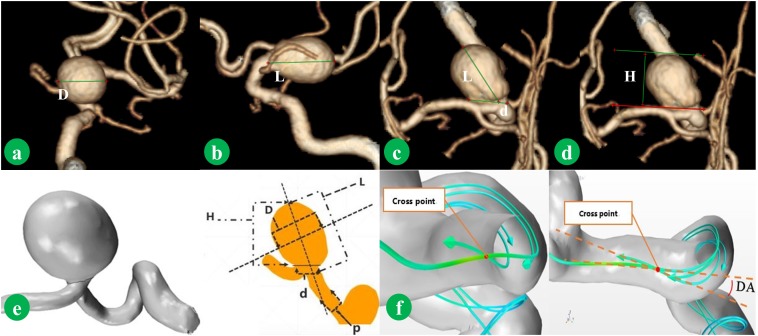
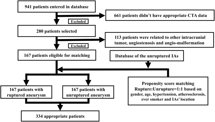
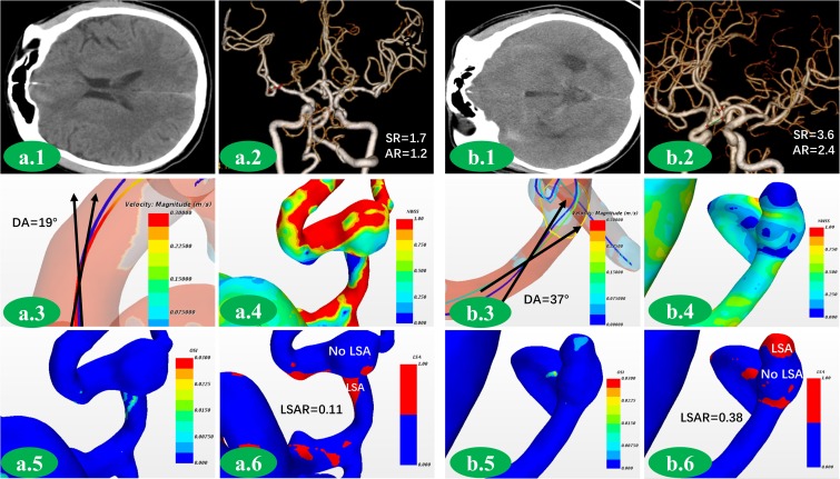
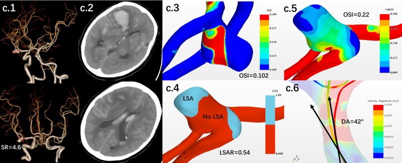
Objective: The aim of the present study is to investigate the potential morphological and hemodynamic risk factors related to intracranial aneurysms (IAs) rupture and establish a system to stratify the risk of IAs rupture to help the clinical decision-making. Methods: Patients admitted to our hospital for single-IAs were selected from January 2012 and January 2018. A propensity score matching was conducted to match patients. The morphological parameters were obtained from high solution CTA images, and the hemodynamic parameters were obtained in accordance with the outcomes of computational fluid dynamics (CFDs) simulation. Differences in the morphologic and hemodynamic parameters were compared. The significant parameters were selected to establish a novel scoring system (Intracranial Aneurysm Rupture Score, IARS). The comparison was drawn between the discriminating accuracy of IARS and the Rupture Resemblance Score (RRS) system to verify the value of IARS. Then, a group of patients with unruptured IAs was stratified into the high risk and low risk groups by IARS and RRS system separately and was followed up for 18-27 months to verify the value of IARS. The outcome of different stratifications was compared. Results: The matching process yielded 167 patients in each group. Differences of statistical significance were found in aneurysm length (p = 0.001), perpendicular height (H) (p < 0.001), aspect ratio (AR) (p < 0.001), size ratio (SR) (p < 0.001), deviated angle (DA) (p < 0.001), normalized average wall shear stress (NWSSa) (p < 0.001), wall shear stress gradient (WSSG) (p < 0.001), low shear area ratio (LSAR) (p = 0.01), and oscillatory shear index (OSI) (p = 0.01). Logistic regression analysis further demonstrated that SR, DA, NWSSa, LSAR, and OSI were the independent risk factors of IAs rupture. SR, DA, LSAR, and OSI were finally selected to establish the IARS. Our present IARS showed a higher discriminating value (AUC 0.81 vs. 0.77) in comparison with the RRS (SR, NWSSa, and OSI). After follow-up, seven patients were subject to IAs rupture. 5/26 in high risk group stratified by IARS, yet 7/57 in high risk group stratified by RRS. The accuracy of IARS was further verified (19.2% vs. 12.3%, AUC for the IARS and the RRS was 0.723 and 0.673, respectively). Conclusion: SR, DA, NWSSa, LSAR, and OSI were considered the independent risk factors of IAs rupture. Our novel IARS showed higher accuracy in discriminating IA rupture in comparison with RRS.

Keywords: computational fluid dynamics; hemodynamic; intracranial aneurysm; morphological; rupture.


Figures

FIGURE 1
FIGURE 1
Morphological parameters were measured from CTA image. D is the maximum diameter of the body (a). L is the maximum distance of the dome from the neck plane (b). d is the average diameter of the neck (c). H is the maximum perpendicular distance of the dome from the neck plane (d). The method of measurement is shown in (e). p is average diameter of the parent artery. Normal vector was combined. The deviated angle (DA), which was between co-velocity and normal vector, was measured (f).
FIGURE 2
FIGURE 2
The selecting flow chart was presented here. A total of 941 patients were enrolled into our study. 167 patients with ruptured IA met our standards in our series. Another 167 patients with unruptured IA were then selected using the propensity score matching. The matching process yielded 167 patients in each group.
FIGURE 3
FIGURE 3
A group of aneurysms with similar match score were presented here. This was an unruptured middle cerebral artery aneurysm (a.1,2). SR was 1.7. AR was 1.2 (a.2). DA was 19°(a.3). NWSS of aneurysm was close to that of parent artery (a.4). There was no area with increasing OSI (a.5). There was no obvious low WSS area in this aneurysm (a.6). This was a ruptured anterior communication artery aneurysm (b.1,2). DA, SR and AR were 37°, 3.6, and 2.4, respectively, which were much higher than unrupture aneurysm (b.2,3). NWSS was lower comparing to parent artery (b.4). Area with obviously increasing OSI could be found (b.5). Large area with low WSS could be found in the dome of aneurysm (b.6).
FIGURE 4
FIGURE 4
The ROC curves of key parameters (DA, SR, LSAR, OSI, and NWSS) were presented here (a,b). The cut-off values for SR, DA, NWSSa, LSAR, OSI, and RRT were 2.3, 35, 0.24, 0.30, 0.008, and 5.3, respectively. The ROC curves of the IARS-SR, IARS-NWSS, and RRS suggested that the AUC of IARS-SR (AUC 0.81) was larger than IARS-NWSS (AUC 0.79) and RRS (AUC 0.77), which suggested that the IARS-SR had higher accuracy to discriminate the ruputre statue (c). For the follow-up group, the distributions of the ruptured aneurysm and unruptured aneurysm in each stratification system were presented (d,e). The IARS has higher AUC (AUC for the IARS and the RRS was 0.723 and 0.673, respectively.
FIGURE 5
FIGURE 5
A female patient with middle cerebral artery aneurysm was followed up by clinical visiting. The patient had hypertension history. Based on the first CTA, SR, DA, LSAR, and OSI were 4.6, 42°, 0.74 and 0.22, respectively (c.1,3,4,5,6). The aneurysm was scored by the IARS as 4, which was stratified as high-risk group. 3.5 months later, the aneurysm was rupture. SAH and hemotoma were confirmed by CT (c.2).

References

    1. Castro M. A. (2013). Understanding the role of hemodynamics in the initiation, progression, rupture, and treatment outcome of cerebral aneurysm from medical image-based computational studies. ISRN Radiol. 2013:602707. 10.5402/2013/602707 - DOI - PMC - PubMed
    1. Cebral J. R., Duan X., Chung B. J., Putman C., Aziz K., Robertson A. M. (2015a). Wall mechanical properties and hemodynamics of unruptured intracranial aneurysms. AJNR Am. J. Neuroradiol. 36 1695–1703. 10.3174/ajnr.A4358 - DOI - PMC - PubMed
    1. Cebral J., Ollikainen E., Chung B. J., Mut F., Sippola V., Jahromi B. R., et al. (2017). Flow conditions in the intracranial aneurysm lumen are associated with inflammation and degenerative changes of the aneurysm wall. AJNR Am. J. Neuroradiol. 38 119–126. 10.3174/ajnr.A4951 - DOI - PMC - PubMed
    1. Cebral J. R., Vazquez M., Sforza D. M., Houzeaux G., Tateshima S., Scrivano E., et al. (2015b). Analysis of hemodynamics and wall mechanics at sites of cerebral aneurysm rupture. J. Neurointerv. Surg. 7 530–536. 10.1136/neurintsurg-2014-011247 - DOI - PubMed
    1. Chen C. J., Moosa S., Ding D., Raper D. M., Burke R. M., Lee C. C., et al. (2016). Infundibular dilations of the posterior communicating arteries: pathogenesis, anatomical variants, aneurysm formation, and subarachnoid hemorrhage. J. Neurointerv. Surg. 8 791–795. 10.1136/neurintsurg-2015-011827 - DOI - PubMed