Review
. 2018 Aug 31:12:274.
doi: 10.3389/fncel.2018.00274. eCollection 2018.

Impairments in Oxidative Glucose Metabolism in Epilepsy and Metabolic Treatments Thereof

Affiliations
Review

Impairments in Oxidative Glucose Metabolism in Epilepsy and Metabolic Treatments Thereof

Tanya McDonald et al. Front Cell Neurosci. .

Abstract

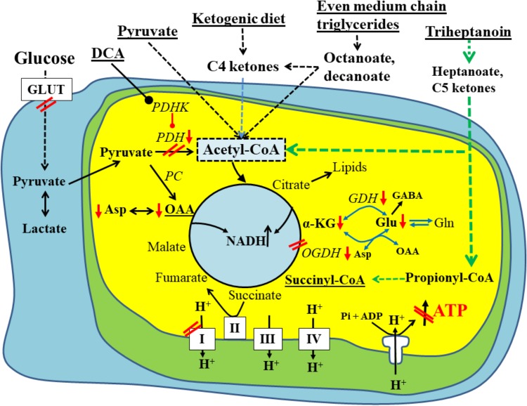
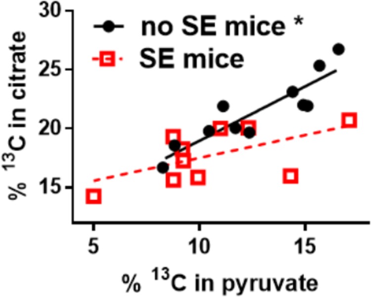
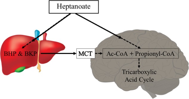
There is mounting evidence that oxidative glucose metabolism is impaired in epilepsy and recent work has further characterized the metabolic mechanisms involved. In healthy people eating a traditional diet, including carbohydrates, fats and protein, the major energy substrate in brain is glucose. Cytosolic glucose metabolism generates small amounts of energy, but oxidative glucose metabolism in the mitochondria generates most ATP, in addition to biosynthetic precursors in cells. Energy is crucial for the brain to signal "normally," while loss of energy can contribute to seizure generation by destabilizing membrane potentials and signaling in the chronic epileptic brain. Here we summarize the known biochemical mechanisms that contribute to the disturbance in oxidative glucose metabolism in epilepsy, including decreases in glucose transport, reduced activity of particular steps in the oxidative metabolism of glucose such as pyruvate dehydrogenase activity, and increased anaplerotic need. This knowledge justifies the use of alternative brain fuels as sources of energy, such as ketones, TCA cycle intermediates and precursors as well as even medium chain fatty acids and triheptanoin.

Keywords: anaplerosis; glucose metabolism; medium chain fatty acids; pilocarpine; temporal lobe epilepsy.


Figures

FIGURE 1
FIGURE 1
Schematic of biochemical impairments found in chronic epileptic tissue and potential treatments thereof. A brain cell under normal physiological conditions uses glucose (left) as the main fuel and produces pyruvate by glycolysis. Pyruvate can be metabolized to lactate in the cytoplasm, if there is an imbalance between glycolysis and TCA cycle activity. Otherwise, pyruvate is taken up by the mitochondria (inner yellow/green area) and is further metabolized by pyruvate dehydrogenase (PDH) to acetyl-CoA, which condenses with oxaloacetate to form citrate, the first intermediate of the TCA cycle. The TCA cycle produces NADH, FADH2, which can be used by the electron transport chain (complexes1-IV on the bottom of diagram) and ATP synthase (complex V) to ultimately produce ATP. Please note that the TCA cycle intermediates are also precursors for lipids and amino acids. The red arrows and double lines indicate reductions in metabolite levels, activities of pathways, enzymes or complexes or glucose uptake, which have been described in epilepsy in either human brains or animal models (see text for explanations). These impairments all limit ATP production by ATP synthase depicted in the diagram as a crossed out arrow in front of ATP in the lower right corner. Several metabolic treatment approaches for epilepsy are underlined and shown on the top and their metabolic effects are indicated. This includes PDH kinase inhibition (such as dichloroacetate) to increase PDH activity as well as direct fuelling with pyruvate, C4 ketone bodies and medium chain fats, which all directly produce acetyl-CoA and do not require PDH for entry into the TCA cycle. Please also note that pyruvate and heptanoate are both providing C4 TCA cycle intermediates, namely oxaloacetate via pyruvate carboxylase (PC) or succinyl-CoA, respectively. This anaplerosis may contribute to TCA cycling as well as biosynthesis of lipids and amino acids from TCA cycle intermediates. α–KG, α-ketoglutarate; GDH, glutamate dehydrogenase; GLUT, glucose transporter; OAA, oxaloacetate; OGDH, oxoglutarate dehydrogenase; PC, pyruvate carboxylase; PDH, pyruvate dehydrogenase; PDHK, PDH kinase.
FIGURE 2
FIGURE 2
Alteration of 13C-pyruvate entry into the TCA cycle in hippocampal tissue of chronically epileptic mice. 13C-glucose was injected (i.p.) into mice that were in the chronic epileptic stage of the pilocarpine SE model (SE mice - red open squares) and control no SE mice, which do not show spontaneous seizures (black circles). 15 min later mice were sacrificed and the hippocampi extracted for LCMS-MS analysis. The % enrichment of 13C-containing citrate was plotted against the % enrichment of 13C-containing pyruvate. A reduction of 13C-carbon entry into the Krebs cycle can be observed in the 17% lower hippocampal % 13C enrichment in citrate in SE vs. no SE mice (p = 0.004, please note that the pyruvate levels were similar). Also, there is a lack of correlation of % 13C enrichment in citrate vs. that of pyruvate in SE mice (slope is not significantly different from zero, p = 0.08), while in no SE mice there is a correlation (p = 0.0002). The lack of correlation indicates that in “epileptic” mice the pyruvate levels do not determine entry into the Krebs cycle, indicating that some other factor is impaired, for example the activity of PDH. Data from (McDonald et al., 2017).
FIGURE 3
FIGURE 3
Heptanoate metabolism. Heptanoate can be taken up by the liver to produce the C5 ketone bodies, β-hydroxypentanoate (BHP) and β-ketopentanoate (BKP), which are then released into blood and taken up by cells through monocarboxylate transporters (MCT). It is also likely that heptanoate directly diffuses into cells, including brain cells. In mitochondria, heptanoate metabolites produce acetyl-CoA and anaplerotic propionyl-CoA, which can enter the TCA cycle directly or after carboxylation as succinyl-CoA, respectively.

References

    1. Akram M. (2014). Citric acid cycle and role of its intermediates in metabolism. Cell Biochem. Biophys. 68 475–478. 10.1007/s12013-013-9750-1 - DOI - PubMed
    1. Alvestad S., Hammer J., Eyjolfsson E., Qu H., Ottersen O. P., Sonnewald U. (2008). Limbic structures show altered glial-neuronal metabolism in the chronic phase of kainate induced epilepsy. Neurochem. Res. 33 257–266. 10.1007/s11064-007-9435-5 - DOI - PubMed
    1. Apelt J., Mehlhorn G., Schliebs R. (1999). Insulin-sensitive GLUT4 glucose transporters are colocalized with GLUT3-expressing cells and demonstrate a chemically distinct neuron-specific localization in rat brain. J. Neurosci. Res. 57 693–705. 10.1002/(SICI)1097-4547(19990901)57:5<693::AID-JNR11>3.0.CO;2-X - DOI - PubMed
    1. Augustin R. (2010). The protein family of glucose transport facilitators: it’s not only about glucose after all. IUBMB Life 62 315–333. 10.1002/iub.315 - DOI - PubMed
    1. Bagga P., Behar K. L., Mason G. F., De Feyter H. M., Rothman D. L., Patel A. B. (2014). Characterization of cerebral glutamine uptake from blood in the mouse brain: implications for metabolic modeling of 13C NMR data. J. Cereb Blood Flow Metab. 34 1666–1672. 10.1038/jcbfm.2014.129 - DOI - PMC - PubMed

LinkOut - more resources