

Identification of Autophagy-Related Gene 7 and Autophagic Cell Death in the Planarian Dugesia japonica
- PMID: 30233400
- PMCID: PMC6131670
- DOI: 10.3389/fphys.2018.01223
Identification of Autophagy-Related Gene 7 and Autophagic Cell Death in the Planarian Dugesia japonica
Abstract
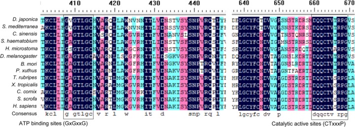
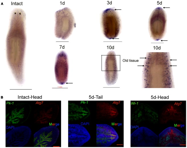
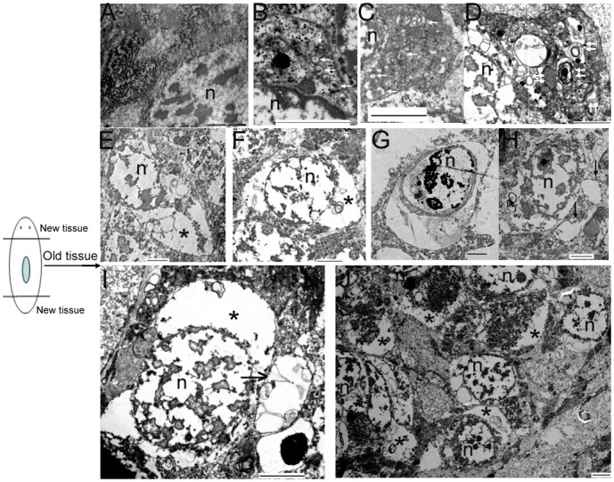
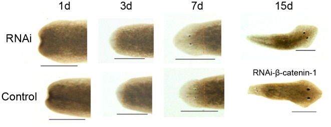
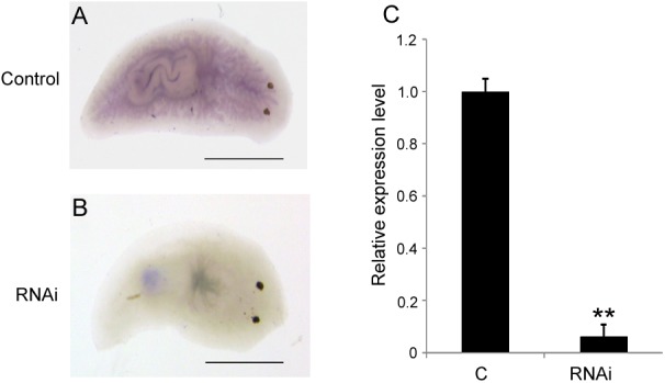
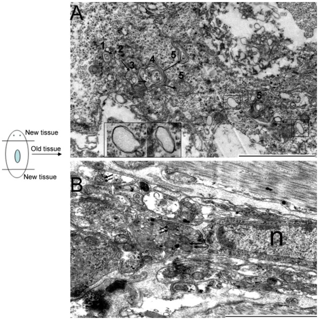
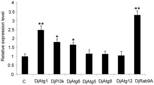
Planarians undergo continuous body size remodeling under starvation or during regeneration. This process likely involves autophagy and autophagic cell death, but this hypothesis is supported by few studies. To test this hypothesis, we cloned and characterized autophagy-related gene 7 (Atg7) from the planarian Dugesia japonica (DjAtg7). The full-length cDNA of DjAtg7 measures 2272 bp and includes a 2082-bp open reading frame encoding 693 amino acids with a molecular weight of 79.06 kDa. The deduced amino acid sequence of DjAtg7 contains a conserved ATP-binding site and a catalytic active site of an E1-like enzyme belonging to the ATG7 superfamily. DjAtg7 transcripts are mainly expressed in intestinal tissues of the intact animals. After amputation, DjAtg7 was highly expressed at the newly regenerated intestinal branch on days 3-7 of regeneration and in the old tissue of the distal intestinal branch on day 10 of regeneration. However, knockdown of DjAtg7 by RNAi did not affect planarian regeneration and did not block autophagosome formation, which indicates that autophagy is more complex than previously expected. Interestingly, TEM clearly confirmed that autophagy and autophagic cell death occurred in the old tissues of the newly regenerated planarians and clearly revealed that the dying cell released vesicles containing cellular cytoplasmic contents into the extracellular space. Therefore, the autophagy and autophagic cell death that occurred in the old tissue not only met the demand for body remodeling but also met the demand for energy supply during planarian regeneration. Collectively, our work contributes to the understanding of autophagy and autophagic cell death in planarian regeneration and body remodeling.
Keywords: DjAtg7; RNAi; autophagic cell death; autophagy; planarian; regeneration.
Figures
Similar articles
-
Autophagy-related DjAtg1-1 plays critical role in planarian regeneration by regulating proliferation and cell death.Cell Tissue Res. 2022 May;388(2):273-286. doi: 10.1007/s00441-022-03591-3. Epub 2022 Feb 2. Cell Tissue Res. 2022. PMID: 35107621
-
Knockdown of Atg1 Impairs Brain Regeneration and Downregulates ECM-Related Genes in the Planarian Dugesia japonica.Mol Neurobiol. 2025 Sep;62(9):11330-11347. doi: 10.1007/s12035-025-04978-3. Epub 2025 Apr 25. Mol Neurobiol. 2025. PMID: 40281299
-
[Functional analysis of autophagy-related gene Atg6 in planarian central nervous system regeneration].Yi Chuan. 2021 Aug 20;43(8):792-801. doi: 10.16288/j.yczz.21-118. Yi Chuan. 2021. PMID: 34413018 Chinese.
-
Autophagy and its role in regeneration and remodeling within invertebrate.Cell Biosci. 2020 Sep 21;10:111. doi: 10.1186/s13578-020-00467-3. eCollection 2020. Cell Biosci. 2020. PMID: 32974004 Free PMC article. Review.
-
Autophagy in invertebrates: insights into development, regeneration and body remodeling.Curr Pharm Des. 2008;14(2):116-25. doi: 10.2174/138161208783378716. Curr Pharm Des. 2008. PMID: 18220823 Review.
Cited by
-
Autophagy-related DjAtg1-1 plays critical role in planarian regeneration by regulating proliferation and cell death.Cell Tissue Res. 2022 May;388(2):273-286. doi: 10.1007/s00441-022-03591-3. Epub 2022 Feb 2. Cell Tissue Res. 2022. PMID: 35107621
-
Djck1α Is Required for Proper Regeneration and Maintenance of the Medial Tissues in Planarians.Cells. 2023 Feb 1;12(3):473. doi: 10.3390/cells12030473. Cells. 2023. PMID: 36766815 Free PMC article.
-
Djhsp60 Is Required for Planarian Regeneration and Homeostasis.Biomolecules. 2022 Jun 9;12(6):808. doi: 10.3390/biom12060808. Biomolecules. 2022. PMID: 35740934 Free PMC article.
-
Autophagy-related Djatg8 is required for remodeling in planarian Dugesia japonica.Biol Open. 2019 Dec 3;8(12):bio045013. doi: 10.1242/bio.045013. Biol Open. 2019. PMID: 31640974 Free PMC article.
-
Knockdown of Atg1 Impairs Brain Regeneration and Downregulates ECM-Related Genes in the Planarian Dugesia japonica.Mol Neurobiol. 2025 Sep;62(9):11330-11347. doi: 10.1007/s12035-025-04978-3. Epub 2025 Apr 25. Mol Neurobiol. 2025. PMID: 40281299
References
-
- Carranza S., Baguna J., Riutort M. (1997). Are the platyhelminthes a monophyletic primitive group? An assessment using 18S rDNA sequences. Mol. Biol. Evol. 14 485–497. - PubMed
-
- do Nascimento L. F., da Silveira L. C., Nisembaum L. G., Colquhoun A., Abe A. S., Mandarim-de-Lacerda C. A., et al. (2016). Morphological and metabolic adjustments in the small intestine to energy demands of growth, storage, and fasting in the first annual cycle of a hibernating lizard (Tupinambis merianae). Comp. Biochem. Physiol. A Mol. Integr. Physiol. 195 55–64. 10.1016/j.cbpa.2016.02.002 - DOI - PubMed
LinkOut - more resources
Full Text Sources
Other Literature Sources
Miscellaneous