. 2018 Sep 4:9:1231.
doi: 10.3389/fphys.2018.01231. eCollection 2018.

An Insect Prostaglandin E2 Synthase Acts in Immunity and Reproduction

Affiliations

An Insect Prostaglandin E2 Synthase Acts in Immunity and Reproduction

Shabbir Ahmed et al. Front Physiol. .

Abstract

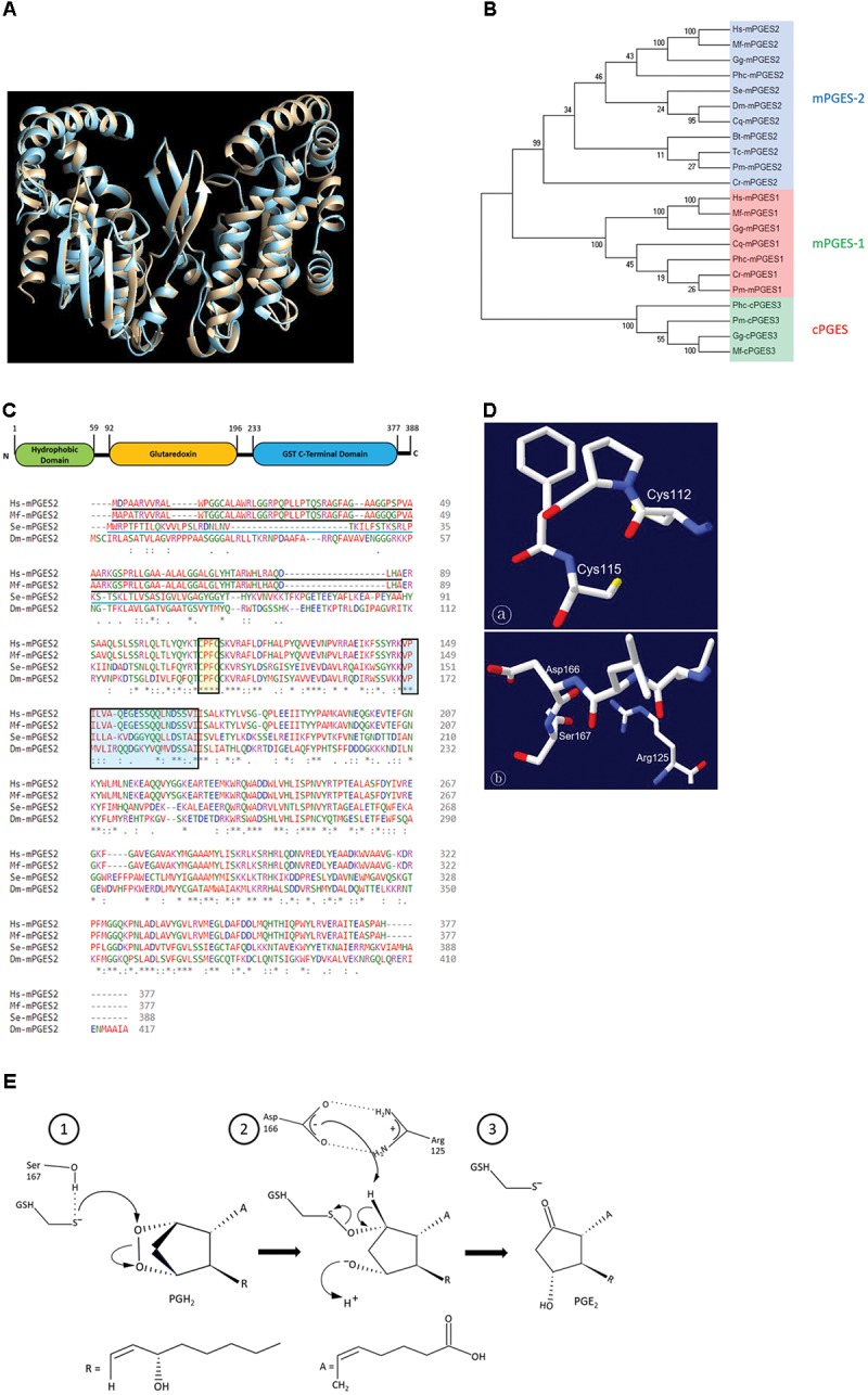
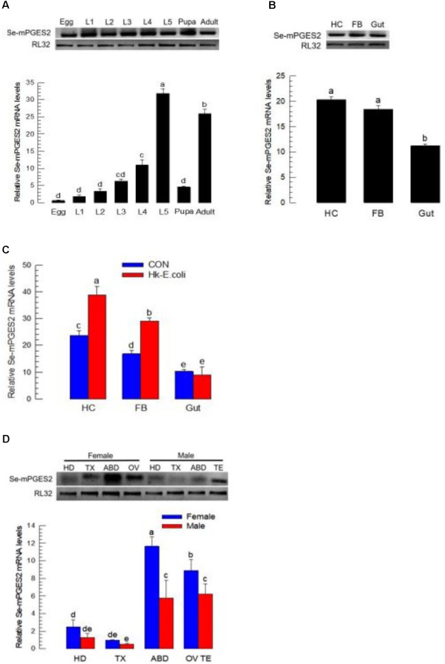
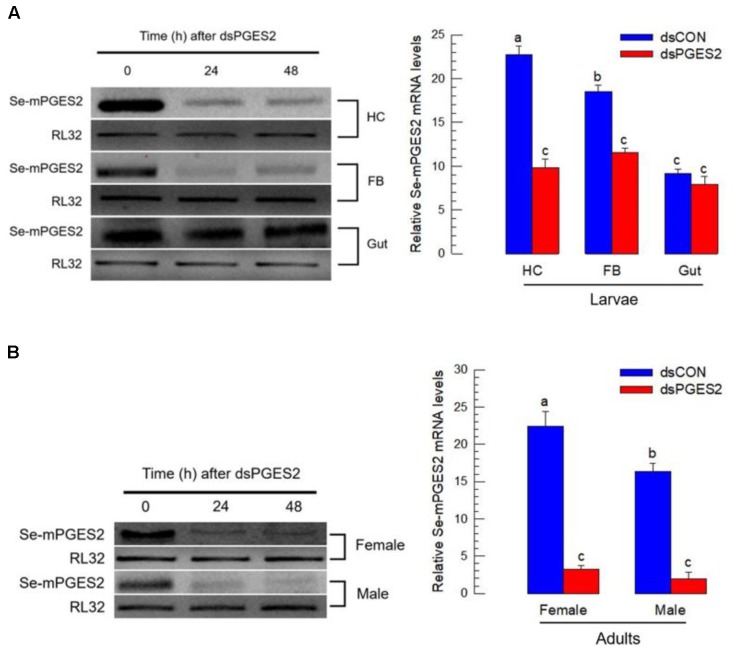
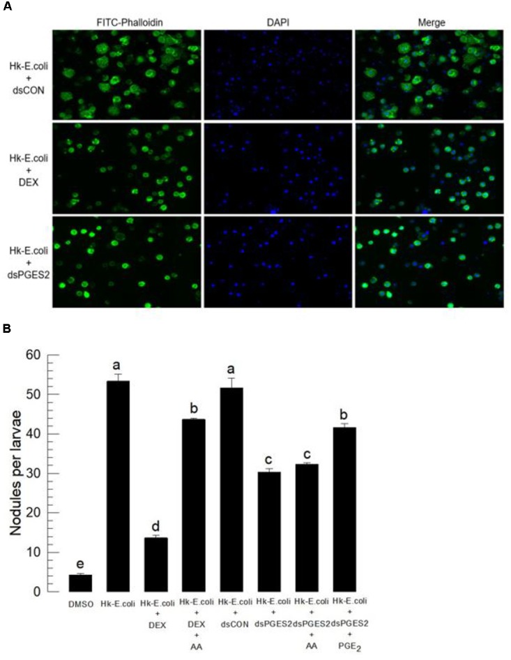
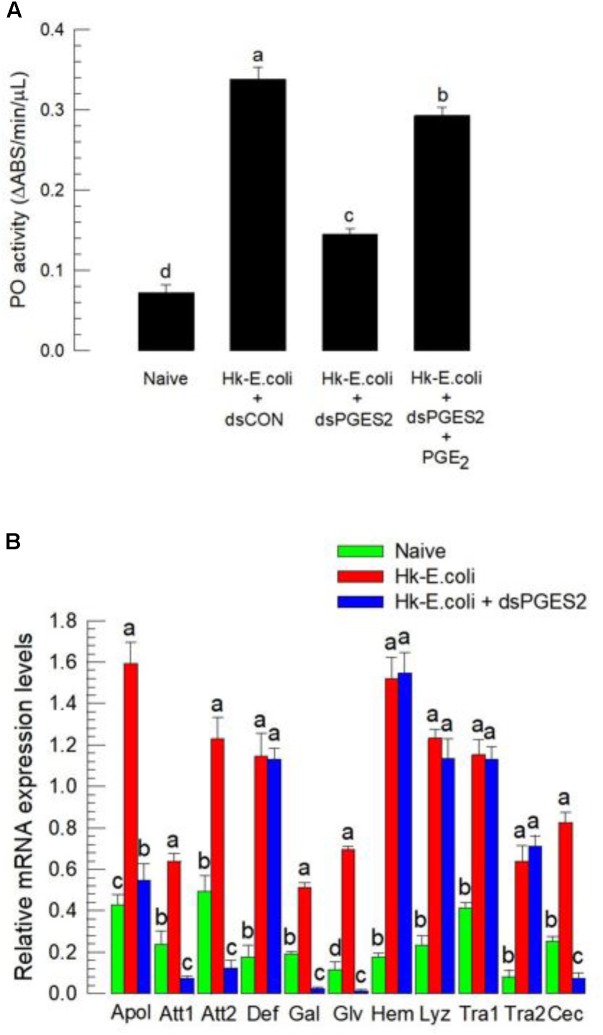
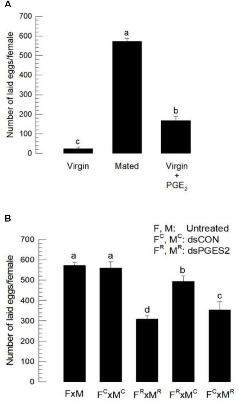
Eicosanoids, oxygenated metabolites of C20 polyunsaturated fatty acids (PUFAs), mediate fundamental physiological processes, including immune reactions and reproduction, in insects. Prostaglandins (PGs) make up one group of eicosanoids, of which PGE2 is a relatively well-known mediator in various insect taxa. While PG biosynthesis has been reported, the specific biosynthetic pathway for PGE2 is not known in insects. Here, we posed the hypothesis that Se-mPGES2 mediates biosynthesis of physiologically active PGE2 through its cognate protein. To test this hypothesis, we interrogated a transcriptome of the lepidopteran insect, Spodoptera exigua, to identify a candidate PGE2 synthase (Se-mPGES2) and analyzed its sequence and expression. Its predicted amino acid sequence contains a consensus thioredoxin homology sequence (Cys-x-x-Cys) responsible for catalytic activity along with an N-terminal membrane-associated hydrophobic domain and C-terminal cytosolic domain. It also shares sequence homology (36.5%) and shares almost overlapping three dimensional structures with a membrane-bound human PGES2 (mPGES2). Se-mPGES2 was expressed in all developmental stages with high peaks during the late larval instar and adult stages. Immune challenge significantly up-regulated its expression levels in hemocytes and fat body. Injecting double-stranded RNA (dsRNA) specific to Se-mPGES2 significantly impaired two cellular immune responses, hemocyte-spreading behavior and nodule formation following bacterial challenge. Humoral immunity was also significantly suppressed, registered as reduced phenoloxidase activity and antimicrobial peptide expression levels. The suppressed immune responses were reversed following PGE2, but not arachidonic acid (AA), treatments. RNAi treatments also reduced the egg-laying behavior of females. Control females mated with the RNAi-treated males led to substantially reduced egg-laying behavior, which was also reversed following PGE2 injections into females. These results strongly bolster our hypothesis that Se-mPGES2 acts in the biosynthesis of PGE2, a crucial biochemical signal mediating immune and reproductive physiology of S. exigua.

Keywords: PGE2; PGES; Spodoptera exigua; eicosanoids; immunity; reproduction.


Figures

FIGURE 1
FIGURE 1
Molecular characteristics of a S. exigua PGES (Se-mPGES2). (A) Structural similarity of Se-mPGES2 to a primate mPGES2. A superimposed view of Macaca fascicularis mPGES-2 crystal structure (gray) and the predicted three-dimensional structure of Se-mPGES2 (blue). Superimposition analysis was performed by UCSF Chimera (https://www.cgl.ucsf.edu/chimera/). (B) A phylogenetic analysis of Se-mPGES2. The analysis was performed using MEGA6. Bootstrapping values were obtained with 1,000 repetitions to support branching and clustering. Amino acid sequences of selected mPGES2 genes were retrieved from GenBank: NP_004869.1 for Homo sapiens (Hs-mPGES1), ADK66307.1 for Gallus gallus (Gg-mPGES1), AAS89037.1 for M. fascicularis (Mf-mPGES-1), ACO11433.1 for Caligus rogercresseyi (Cr-mPGES1), AFJ11395.1 for Penaeus monodon (Pm-mPGES-1), XP_001863047.1 for Culex quinquefasciatus (Cq-mPGES1), EEB10983.1 for Pediculus humanus corporis (Phc-mPGES1), NP_079348.1 for H. sapiens (Hs-mPGES2), XP_415498.1 G. gallus (Gg-mPGES2), BAB01608.1 for M. fascicularis (Mf-mPGES2), ACO11658.1 for C. rogercresseyi (Cr-mPGES2), NP_524116.2 for D. melanogaster (Dm-mPGES-2), XP_002432321.1 for P. humanus corporis (Phc-mPGES2), XP_001868980.1 for C. quinquefasciatus (Cq-mPGES-2), XP_003403370.3 for Bombus terrestris (Bt-mPGES2), XP_973652.1 for Tribolium castaneum (Tc-mPGES2), AFJ11396.1 for P. monodon (Pm-mPGES2), Q90955.1 for G. gallus (Gg-cPGES3), AAS89038.1 for M. fascicularis (Mf-cPGES3), XP_002430923.1 for P. humanus corporis (Phc-cPGES3), and AFJ11394.1 for P. monodon (Pm-cPGES3). (C) Domain analysis of Se-mPGES2. The domains of Se-mPGES2 were predicted using Pfam (http://pfam.xfam.org) and Prosite (https://prosite.expasy.org/). Se-mPGES2 was aligned with the deduced amino acid sequences of H. sapiens, M. fascicularis, and Drosophila melanogaster mPGES2. Identical amino acids were marked with asterisks while similar amino acids were denoted with colons. The N-terminal hydrophobic domain was predicted and underlined with a solid line. The Cys-Pro-Phe-Cys motif and predicted GSH-binding motif were boxed. (D) A model structure of the Cys-Pro-Phe-Cys motif and GSH-binding motif. Swiss-PDB Viewer (http://spdv.vital-it.ch/) was used for detection of protein motifs and active sites. (E) A proposed mechanism of the catalytic activity of Se-mPGES2 against isomerization of PGH2 into PGE2.
FIGURE 2
FIGURE 2
Expression profile of Se-mPGES2. (A) Expression patterns in different developmental stages: egg, first to fifth instar larvae (“L1–L5”), pupa, and adult. (B) Expression patterns in indicated tissues of L5 larvae: hemocyte (“HC”), fat body (“FB”), and midgut (“Gut”). (C) Induction of Se-mPGES2 in response to bacterial challenge. Heat-killed E. coli was injected into L5 larvae and incubated for 12 h at 25°C. (D) Expression patterns in indicated body parts of adults: head (“HD”), thorax (“TX”), abdomen (“ABD”), ovary (“OV”), and testis (“TE”). A ribosomal gene, RL32, was used as reference gene. Each treatment was replicated three times with independent tissue preparations. Different letters indicate significant differences among means at Type I error = 0.05 (LSD test).
FIGURE 3
FIGURE 3
RNA interference (RNAi) of Se-mPGES2 in larvae and adults. One mg of gene-specific dsRNA (“dsPGES2”) was injected into L5D1 or 5 days old pupae. A viral gene, CpBV302, was used as a control dsRNA (“dsCON”). (A) Effect of RNAi on Se-mPGES2 expression in indicated tissues of L5 larvae: hemocyte (“HC”), fat body (“FB”) and midgut (“Gut”). (B) Effect of RNAi on Se-mPGES2 expression in male and female adults. Each treatment was independently replicated three times. Different letters indicate significant differences among means at Type I error = 0.05 (LSD test).
FIGURE 4
FIGURE 4
Bioassay of a physiological role of Se-mPGES2 in cellular immunity after dsRNA treatments, performed as described in Figure 3. For a bacterial challenge, heat-killed (HK) E. coli (5.4 × 10⁴ cells per larva) was injected into larvae at 48 h after dsRNA treatment. (A) Inhibitory effect of dsSe-mPGES2 against F-actin growth in response to bacterial challenge. At 4 h PI, hemocytes were observed under a fluorescence microscope at 40× magnification. Hemocytic F-actin filaments were specifically recognized by FITC-tagged phalloidin (green) while nucleus was stained with DAPI (blue). A PLA2 inhibitor, dexamethasone (“DEX”), was used along with bacteria. (B) Inhibitory effect of dsSe-mPGES2 against hemocyte nodule formation in response to the bacterial challenge. At 8 h PI, numbers of nodules were assessed. Each treatment was independently replicated five times. Different letters indicate significant differences among means at Type I error = 0.05 (LSD test).
FIGURE 5
FIGURE 5
The influence of dsSe-mPGES2 treatments on humoral immunity. dsRNA injected as described in Figure 3. For a bacterial challenge, heat-killed (HK) E. coli (5.4 × 10⁴ cells per larva) was injected into larvae at 48 h after dsRNA treatment. (A) Inhibitory effect of dsSe-mPGES2 on phenoloxidase (PO) activity in response to bacterial challenge. At 8 h PI of the bacteria, PO activity was measured. PGE2 (1 μg per larva) was injected along with the bacteria. (B) Inhibitory effect of Se-mPGES2 RNAi against the expression of 11 antimicrobial peptide (AMP) genes: apolipophorin III (“Apol”), attacin 1 (“Att1”), attacin 2 (“Att2”), defensin (“Def”), gallerimycin (“Gal”), gloverin (“Glv”), hemolin (“Hem”), lysozyme (“Lyz”), transferrin 1 (“Tra1”), transferrin 2 (“Tra2”), and cecropin (“Cec”). RL32 was used as an internal control. Each treatment was replicated three times. Different letters indicate significant differences among means at Type I error = 0.05 (LSD test).
FIGURE 6
FIGURE 6
The influence of dsSe-mPGES2 treatments on reproduction. dsRNA was injected as described in Figure 3 into 5 days old male (“M”) or female (“F”) pupae. A viral gene, CpBV302, was used as a control dsRNA (“dsCON”). (A) Stimulatory effect of PGE2 on egg-laying behavior. Total number of laid eggs by virgin (without male) or mated (with males in 1:1 sex ratio) was counted for 3 days after adult emergence. PGE2 (10 μg per adult) was injected to virgin females at the day of adult emergence. (B) Inhibitory effect of dsSe-mPGES2 treatments on egg-laying behavior. Each treatment consisted of three replicates. Each replicate used 10 females and/or 10 males. Different letters indicate significant differences among means at Type I error = 0.05 (LSD test).

Similar articles

Cited by

References

    1. Bichon A., Boukhatem N., Gay P., Dru P., Terzian H., Petitjean A. M., et al. (2001). Genetic and molecular features of Su(P), a gene that interacts with ref(2)P in male fertility of Drosophila melanogaster. Mol. Genet. Genomics 265 354–361. - PubMed
    1. Bustin S. A., Benes V., Garson J. A., Hellemans J., Huggett J., Kubista M., et al. (2009). The MIQE guidelines: minimum information for publication of quantitative real-time PCR experiments. Clin. Chem. 55 611–622. 10.1373/clinchem.2008.112797 - DOI - PubMed
    1. Cerenius L., Söderhäll K. (2004). The prophenoloxidase-activating system in invertebrates. Immunol. Rev. 198 116–126. 10.1111/j.0105-2896.2004.00116.x - DOI - PubMed
    1. Dalton T. (1977). The effect of prostaglandin E1 on cyclic AMP production in the salivary glands of Calliphora erythrocephala. Experientia 33 1329–1330. 10.1007/BF01920162 - DOI - PubMed
    1. Destephano D. B., Brady U. E., Lovins R. E. (1974). Synthesis of prostaglandin by reproductive tissue of the male house cricket, Acheta domesticus. Prostaglandins 6 71–79. 10.1016/S0090-6980(74)80042-0 - DOI - PubMed

LinkOut - more resources