

Remodeling of Retinal Architecture in Diabetic Retinopathy: Disruption of Ocular Physiology and Visual Functions by Inflammatory Gene Products and Pyroptosis
- PMID: 30233418
- PMCID: PMC6134046
- DOI: 10.3389/fphys.2018.01268
Remodeling of Retinal Architecture in Diabetic Retinopathy: Disruption of Ocular Physiology and Visual Functions by Inflammatory Gene Products and Pyroptosis
Abstract
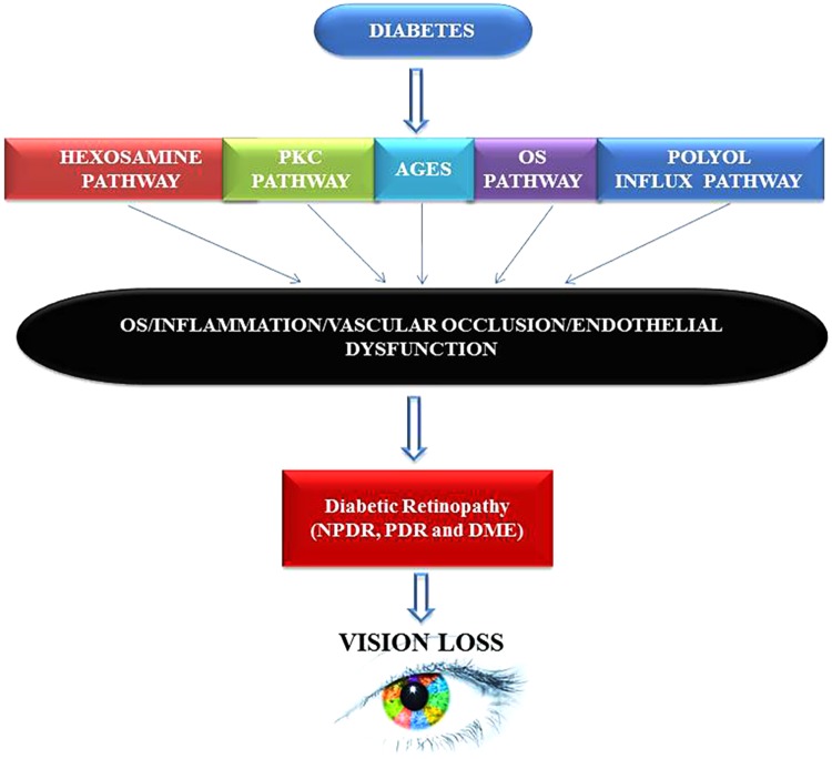
Diabetic patients suffer from a host of physiological abnormalities beyond just those of glucose metabolism. These abnormalities often lead to systemic inflammation via modulation of several inflammation-related genes, their respective gene products, homocysteine metabolism, and pyroptosis. The very nature of this homeostatic disruption re-sets the overall physiology of diabetics via upregulation of immune responses, enhanced retinal neovascularization, upregulation of epigenetic events, and disturbances in cells' redox regulatory system. This altered pathophysiological milieu can lead to the development of diabetic retinopathy (DR), a debilitating vision-threatening eye condition with microvascular complications. DR is the most prevalent cause of irreversible blindness in the working-age adults throughout the world as it can lead to severe structural and functional remodeling of the retina, decreasing vision and thus diminishing the quality of life. In this manuscript, we attempt to summarize recent developments and new insights to explore the very nature of this intertwined crosstalk between components of the immune system and their metabolic orchestrations to elucidate the pathophysiology of DR. Understanding the multifaceted nature of the cellular and molecular factors that are involved in DR could reveal new targets for effective diagnostics, therapeutics, prognostics, preventive tools, and finally strategies to combat the development and progression of DR in susceptible subjects.
Keywords: chemokines; cytokines; diabetic retinopathy; epigenomics; homocysteine; inflammation; pyroptosis; signaling pathways.
Figures
Similar articles
-
Elucidating glial responses to products of diabetes-associated systemic dyshomeostasis.Prog Retin Eye Res. 2023 May;94:101151. doi: 10.1016/j.preteyeres.2022.101151. Epub 2023 Apr 5. Prog Retin Eye Res. 2023. PMID: 37028118 Free PMC article. Review.
-
Targeting the NLRP3 inflammasome in diabetic retinopathy: From pathogenesis to therapeutic strategies.Biochem Pharmacol. 2023 Jun;212:115569. doi: 10.1016/j.bcp.2023.115569. Epub 2023 Apr 25. Biochem Pharmacol. 2023. PMID: 37100255 Review.
-
The mechanisms of NLRP3 inflammasome/pyroptosis activation and their role in diabetic retinopathy.Front Immunol. 2023 Apr 25;14:1151185. doi: 10.3389/fimmu.2023.1151185. eCollection 2023. Front Immunol. 2023. PMID: 37180116 Free PMC article. Review.
-
Mechanistic Insights into Pathological Changes in the Diabetic Retina: Implications for Targeting Diabetic Retinopathy.Am J Pathol. 2017 Jan;187(1):9-19. doi: 10.1016/j.ajpath.2016.08.022. Epub 2016 Nov 12. Am J Pathol. 2017. PMID: 27846381 Free PMC article. Review.
-
Reduction of Glut1 in the Neural Retina But Not the RPE Alleviates Polyol Accumulation and Normalizes Early Characteristics of Diabetic Retinopathy.J Neurosci. 2021 Apr 7;41(14):3275-3299. doi: 10.1523/JNEUROSCI.2010-20.2021. Epub 2021 Feb 23. J Neurosci. 2021. PMID: 33622781 Free PMC article.
Cited by
-
Restoration of skeletal muscle homeostasis by hydrogen sulfide during hyperhomocysteinemia-mediated oxidative/ER stress condition 1.Can J Physiol Pharmacol. 2019 Jun;97(6):441-456. doi: 10.1139/cjpp-2018-0501. Epub 2018 Nov 13. Can J Physiol Pharmacol. 2019. PMID: 30422673 Free PMC article. Review.
-
Hyaluronic-Coated Albumin Nanoparticles for the Non-Invasive Delivery of Apatinib in Diabetic Retinopathy.Int J Nanomedicine. 2021 Jul 2;16:4481-4494. doi: 10.2147/IJN.S316564. eCollection 2021. Int J Nanomedicine. 2021. PMID: 34239300 Free PMC article.
-
N-Methyl-D-aspartate receptor activation, novel mechanism of homocysteine-induced blood-retinal barrier dysfunction.J Mol Med (Berl). 2021 Jan;99(1):119-130. doi: 10.1007/s00109-020-02000-y. Epub 2020 Nov 6. J Mol Med (Berl). 2021. PMID: 33159240 Free PMC article.
-
PSAT1 is upregulated by METTL3 to attenuate high glucose-induced retinal pigment epithelial cell apoptosis and oxidative stress.Diagn Pathol. 2024 Oct 15;19(1):138. doi: 10.1186/s13000-024-01556-4. Diagn Pathol. 2024. PMID: 39407268 Free PMC article.
-
Inhibition of the polyol pathway by Ducrosia anethifolia extract: plausible implications for diabetic retinopathy treatment.Front Pharmacol. 2024 Dec 23;15:1513967. doi: 10.3389/fphar.2024.1513967. eCollection 2024. Front Pharmacol. 2024. PMID: 39764459 Free PMC article.
References
-
- Aiello L. P., Bursell S. E., Clermont A., Duh E., Ishii H., Takagi C., et al. (1997). Vascular endothelial growth factor-induced retinal permeability is mediated by protein kinase C in vivo and suppressed by an orally effective beta-isoform-selective inhibitor. Diabetes Metab. Res. Rev. 46 1473–1480. - PubMed
Publication types
Grants and funding
LinkOut - more resources
Full Text Sources
Other Literature Sources