. 2018 Sep 4:9:2081.
doi: 10.3389/fmicb.2018.02081. eCollection 2018.

Porphyromonas gingivalis Promotes 4-Nitroquinoline-1-Oxide-Induced Oral Carcinogenesis With an Alteration of Fatty Acid Metabolism

Affiliations

Porphyromonas gingivalis Promotes 4-Nitroquinoline-1-Oxide-Induced Oral Carcinogenesis With an Alteration of Fatty Acid Metabolism

Jia-Shun Wu et al. Front Microbiol. .

Abstract

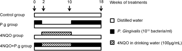
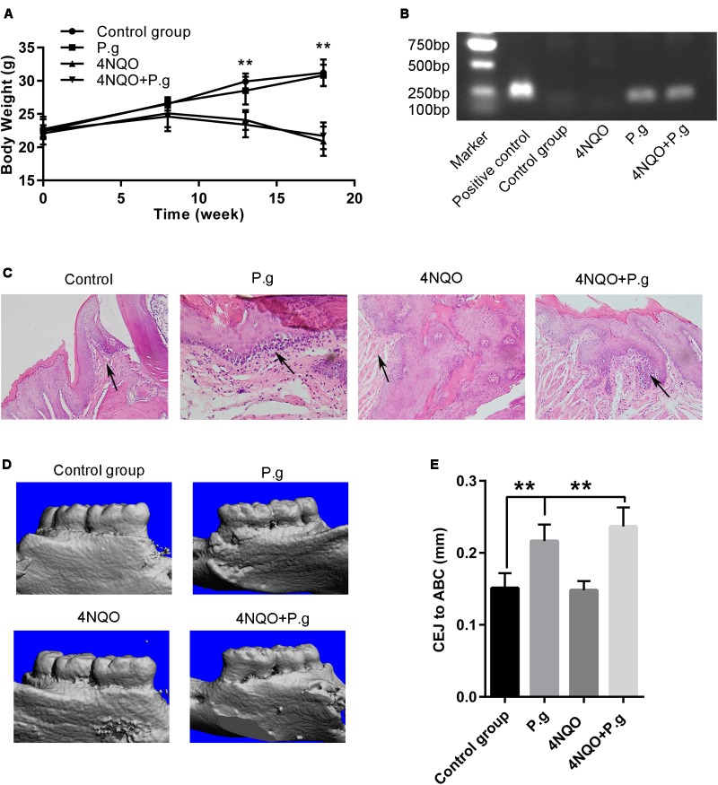
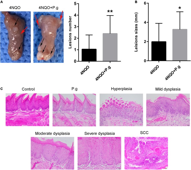
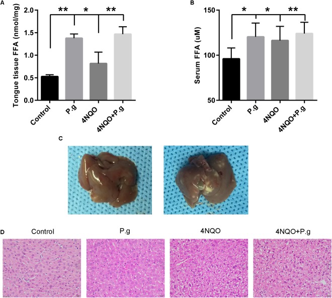
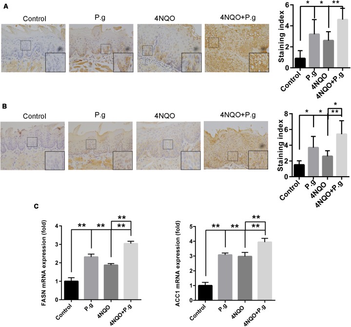
Microbiota has been widely considered to play a critical role in human carcinogenesis. Human papilloma virus, hepatitis B and C virus, and Helicobacter pylori are implicated in the pathogenesis of cancer of uterine cervix, liver, and stomach, respectively. However, whether Porphyromonas gingivalis (P. gingivalis), a common Gram negative oral bacteria, is associated with oral carcinogenesis still remains unclear and its underlying mechanism needs to be addressed. Here, we established a combined experimental system of 4NQO-induced oral carcinoma model and chronic periodontitis model and investigated the effects of P. gingivalis infection on oral carcinogenesis and fatty acid metabolism during oral carcinogenesis. The data showed that in this animal model, P. gingivalis infection induced mice periodontitis, increased the tongue lesion size and multiplicity of each mouse and promoted oral cancer development. P. gingivalis treatment significantly increased the level of free fatty acids and altered the fatty acid profile in tongue tissues and the serum of mice. And P. gingivalis induced the formation of fatty liver of the mice. Besides, immunohistochemical analysis and qRT-PCR showed that the expression of fatty-acid synthase and acetyl-CoA carboxylase 1 were increased in the tongue and liver tissues of 4NQO-treated mice infected with P. gingivalis. These results showed that P. gingivalis promoted oral carcinogenesis and aggravated disturbance of fatty acid metabolism, indicating a close association among P. gingivalis, lipid metabolic and oral carcinogenesis.

Keywords: 4-nitroquinoline-1-oxide; Porphyromonas gingivalis; fatty acid synthases; fatty acids; mouse models; oral squamous cell carcinoma.


Figures

FIGURE 1
FIGURE 1
Diagram of the experimental protocol. Oral administration of P. gingivalis was given to the mice in P.g and 4NQO + P.g group in the period of 0–2 weeks and 10–18 weeks. And the mice in 4NQO and 4NQO + P.g group were exposed to 4NQO treatment in the period of 2–10 weeks. The control mice were treated with vehicle alone.
FIGURE 2
FIGURE 2
Oral administration of P. gingivalis induced periodontitis in the mice. (A) The body weights of animals in control, P.g, 4NQO, and 4NQO + P.g group. The data represent the mean ± SEM of 10 animals per group at the indicated times; ∗∗P < 0.01. (B) Representative results of colony PCR for detection of P. gingivalis in the mice oral cavity. (C) Histochemical analysis of gingival tissues of mice each group. The histological images of gingival tissues in P.g and in 4NQO + P.g group had more inflammatory cell infiltration compared with 4NQO and control group. All images were taken at 400× magnification. (D) Representative results of micro-CT analysis of maxillary hemi-jaws in mice from each group. The levels of alveolar bone crest (ABC) in mice of control and 4NQO group were at the cement-enamel junction (CEJ), whereas severe bone absorption was observed in mice of P.g and 4NQO + P.g group. (E) The quantitative data of alveolar bone loss was measured as distance from the CEJ to the ABC. The columns represent the mean ± SEM of 10 animals per group; ∗∗P < 0.001.
FIGURE 3
FIGURE 3
Effect of P. gingivalis infection on tongue carcinogenesis in 4NQO-treated mice. (A) Representative imagines tongue tissues and the quantitative data of lesion number per mice in 4NQO and 4NQO + P.g group were shown. Black arrows indicate tumors; red arrows indicate dysplasia. The columns represent the mean ± SEM of 10 animals per group; ∗∗P < 0.01. (B) The average of lesion sizes in mice of 4NQO and 4NQO + P.g group. The columns represent the mean ± SEM of 10 animals per group; P < 0.05. (C) Representative HE sections of pathology, including hyperplasia, mild dysplasia, moderate dysplasia, severe dysplasia, and carcinoma. All images were taken at 400× magnification.
FIGURE 4
FIGURE 4
Effect of P. gingivalis infection on FFAs levels in tongue tissues and serum in 4NQO-treated mice. The levels of FFAs in tongue tissues (A) and serum (B) in different group mice were shown. The columns represent the mean ± SEM of 10 animals per group; P < 0.05; ∗∗P < 0.001. (C) Representative imagines of mice liver in non-P. gingivalis-treatment group (left panel) and P. gingivalis -treatment group (right panel).The liver of P. gingivalis-treated mice (P.g and 4NQO + P.g group) exhibited an appearance of fatty liver with yellow-white spots compared with uniform and normal appearance of the control group. (D) Representative HE sections of liver tissues from mice in each experimental group. The liver of 4NQO-treated mice exhibited drug mediated liver injury with characteristics of hepatocyte ballooning and mild microvesicular steatosis. Interestingly, the liver of P. gingivalis-treated mice showed some pathological changes of fatty liver including micro- and macrovesicular steatosis. All images were taken at 200× magnification.
FIGURE 5
FIGURE 5
Effect of P. gingivalis infection on expression of FASN and ACC1 in tongue tissues of 4NQO-treated mice. IHC analyses were performed in tongue tissues from each experimental group following the procedures described in Section “Materials and Methods.” Representative photographs for FASN (A) and ACC1 (B) were showed and at least five mice from each experimental group with two images per mouse were quantified. These results revealed that greater levels of FASN and ACC1 in 4NQO + P.g group than in 4NQO group. All images were taken at 400× magnification. Each column represents the mean ± SD; P < 0.05; ∗∗P < 0.001. (C) Graphics of mRNA expression levels of FASN and ACC1 by RT-qPCR for samples from each experimental group. The columns represent the mean ± SEM of five animals per group; ∗∗P < 0.001.

References

    1. Agostini M., Almeida L. Y., Bastos D. C., Ortega R. M., Moreira F. S., Seguin F., et al. (2014). The fatty acid synthase inhibitor orlistat reduces the growth and metastasis of orthotopic tongue oral squamous cell carcinomas. Mol. Cancer Ther. 13 585–595. 10.1158/1535-7163.mct-12-1136 - DOI - PubMed
    1. Agostini M., Silva S. D., Zecchin K. G., Coletta R. D., Jorge J., Loda M., et al. (2004). Fatty acid synthase is required for the proliferation of human oral squamous carcinoma cells. Oral Oncol. 40 728–735. 10.1016/j.oraloncology.2004.01.011 - DOI - PubMed
    1. Ahn J., Segers S., Hayes R. B. (2012). Periodontal disease, Porphyromonas gingivalis serum antibody levels and orodigestive cancer mortality. Carcinogenesis 33 1055–1058. 10.1093/carcin/bgs112 - DOI - PMC - PubMed
    1. Askari M., Darabi M., Zare Mahmudabadi R., Oboodiat M., Fayezi S., Mostakhdemin Hosseini Z., et al. (2015). Tissue fatty acid composition and secretory phospholipase-A2 activity in oral squamous cell carcinoma. Clin. Transl. Oncol. 17 378–383. 10.1007/s12094-014-1242-2 - DOI - PubMed
    1. Barber G. N. (2015). STING: infection, inflammation and cancer. Nat. Rev. Immunol. 15 760–770. 10.1038/nri3921 - DOI - PMC - PubMed

LinkOut - more resources