Review
. 2018 Sep 4:9:2085.
doi: 10.3389/fmicb.2018.02085. eCollection 2018.

Circular and Leaderless Bacteriocins: Biosynthesis, Mode of Action, Applications, and Prospects

Affiliations
Review

Circular and Leaderless Bacteriocins: Biosynthesis, Mode of Action, Applications, and Prospects

Rodney H Perez et al. Front Microbiol. .

Abstract

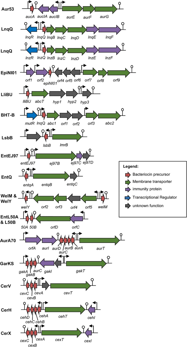
Bacteriocins are a huge family of ribosomally synthesized peptides known to exhibit a range of bioactivities, most predominantly antibacterial activities. Bacteriocins from lactic acid bacteria are of particular interest due to the latter's association to food fermentation and the general notion of them to be safe. Among the family of bacteriocins, the groups known as circular bacteriocins and leaderless bacteriocins are gaining more attention due to their enormous potential for industrial application. Circular bacteriocins and leaderless bacteriocins, arguably the least understood groups of bacteriocins, possess distinctively peculiar characteristics in their structures and biosynthetic mechanisms. Circular bacteriocins have N-to-C- terminal covalent linkage forming a structurally distinct circular peptide backbone. The circular nature of their structures provides them superior stability against various stresses compared to most linear bacteriocins. The molecular mechanism of their biosynthesis, albeit has remained poorly understood, is believed to possesses huge application prospect as it can serve as scaffold in bioengineering other biologically important peptides. On the other hand, while most bacteriocins are synthesized as inactive precursor peptides, which possess an N-terminal leader peptide attached to a C-terminal propeptide, leaderless bacteriocins are atypical as they do not have an N-terminal leader peptide, hence the name. Leaderless bacteriocins are active right after translation as they do not undergo any post-translational processing common to other groups of bacteriocins. This "simplicity" in the biosynthesis of leaderless bacteriocins offers a huge commercial potential as scale-up production systems are considerably easier to assemble. In this review, we summarize the current studies of both circular and leaderless bacteriocins, highlighting the progress in understanding their biosynthesis, mode of action, application and their prospects.

Keywords: bacteriocin biosynthesis; bacteriocins; circular bacteriocins; lactic acid bacteria; leaderless bacteriocins; mode of action.


Figures

FIGURE 1
FIGURE 1
Primary structures of circular bacteriocin precursor peptides. Amino acid positions are numbered and the point of cleavage between the leader peptide and the core peptide is indicated by an arrow. Enterocin AS-48 (AS-48), pumilarin (Pmln), amylocyclicin (Acn), enterocin NKR-5-3B (Ent53B), gassericin A (GaaA), butyrivibriocin AR10 (BviA), acidocin B (AciB), and plantaricyclin A (PlnA) are members of the circular bacteriocins with long leader sequence, whereas circularin A (CirA), uberolysin A (UblA), carnocyclin A (CclA), leucocyclicin Q (LcyQ), lactocyclicin Q (LycQ), and garvicin ML (GarML) belong to circular bacteriocins with short leader sequence. Sub-grouping based on their biochemical features are also indicated as groups i and ii. Circular bacteriocins belonging to group ii showed high sequence homology. Whereas overall sequence comparison of group i bacteriocins showed very limited sequence homology, however, pairwise comparison between, AS-48 and Pmln, Acn and Ent53B, and LcyQ and LycQ showed very high sequence identity. Intensity (black to gray) of the highlighted residue indicates strong conservation of their residues.
FIGURE 2
FIGURE 2
Biosynthetic gene cluster of circular bacteriocins. Genes are color coded based on their known and putative functions as indicated. Abbreviation of bacteriocins are shown in the legend of Figure 1. The bent arrows and lollipop symbols represent putative promoters and putative terminators, respectively. Figures are not drawn in scale.
FIGURE 3
FIGURE 3
Primary structures of leaderless bacteriocins. Leaderless bacteriocins are clustered based on the number of their component peptides; single-peptide, two-peptide, and multi-peptide. Single peptide group is composed of aureocin A53 (Aur53), lacticins Q and Z (LnqQ and Z), epidermicin NI01 (EpiNI01), lactolisterin BU (LliBU), BHT-B, LsbB, enterocins EJ97, K1, and Q (EntEJ97, EntK1, and EntQ, respectively), and weissellicins M and Y (WelM and Y). Two-peptide group is composed of enterocins L50 (L50A and B) and enterocins MR10 (EntMR10A and B). Whereas multi-peptide group is composed of aureocin A70 (AurA, B, C, and D), garvicin KS (GakA, B, and C), cereucin V (Cev A, B, and C), cereucin X (cexA, B, and C), and cereucin H (CehA, B, C, and D). Sub-clustering based on their sequence homology is also indicated; aur53-like, LsbB-like, and entL50-like. Intensity (black to gray) of the highlighted residue indicates strong conservation of their residues.
FIGURE 4
FIGURE 4
Biosynthetic gene cluster of leaderless bacteriocins. Genes are color coded based on their known and putative functions as indicated. Abbreviation of bacteriocins are shown in the legend of Figure 3. The bent arrows and lollipop symbols represent putative promoters and putative terminators, respectively. Figures are not drawn in scale.
FIGURE 5
FIGURE 5
Cartoon representation of the structures of the representative sub-groups of circular and leaderless bacteriocins. (A) enterocin NKR-5-3B represents the saposin fold exhibiting circular bacteriocins or group i, (B) acidocin B represents the helical bundle structure exhibiting circular bacteriocins or group ii, (C) lacticin Q represents the saposin fold exhibiting single peptide leaderless bacteriocins, (D) LsbB represents the single helical structure exhibiting single peptide leaderless bacteriocins, and (E) enterocin 7, composed of enterocin 7A and 7B, represents the saposin fold exhibiting two-peptide leaderless bacteriocins. N- and C-terminal ligation site of the circular bacteriocins is indicated by arrow whereas the terminal ends of leaderless bacteriocins are indicated by letters N and C. In order to depict directionality, helices of each bacteriocins are numbered accordingly. Images are rendered using PyMol Molecular Graphics System.
FIGURE 6
FIGURE 6
Sequence and structure alignment of representative peptides of saposin fold exhibiting bacteriocins; enterocin NKR-5-3B (Ent53B), lacticin Q (LnqQ), enterocin 7 (Ent7A and 7B), saposin and saposin-like peptide. (A) Sequence alignment highlighting the helical and coil regions of these peptides as represented by the cylinder and line symbols, respectively, (B) pair-wise superimposition of the structure of saposin C (SAPC; blue) with the structures of NK-lysin (NKL; red), enterocin NKR-5-3B (Ent53B; green), lacticin Q (LnqQ; yellow–green), enterocin 7 (Ent7A; cyan and 7B; pink). The helices are numbered accordingly with SAPC as the reference. The N-termini of the peptides and the ligation site of Ent53B are indicated by the letter N and an arrow, respectively. In the case of Ent53B the region in the “fifth helix” is repeated in the first helix (amino acid sequence are colored red) due to the N- and C-terminal ligation. Images are generated using PyMol Molecular Graphics System.
FIGURE 7
FIGURE 7
Hydrophobic surface maps of representative sub-groups of circular and leaderless bacteriocins. Enterocin NKR-5-3B and acidocin B represent group i and group ii circular bacteriocins, respectively. Lacticin Q and LsbB represent saposin fold exhibiting and single helical structure exhibiting single peptide leaderless bacteriocins, respectively. Enterocin 7 (Ent 7A and Ent 7B) represents the saposin fold exhibiting two-peptide leaderless bacteriocins. Red indicates hydrophobic residues and white represents hydrophilic resides. The color intensity indicates the hydrophobicity or hydrophilicity of the residues. Images are generated using PyMol Molecular Graphics System.

References

    1. Abengózar M., Á Cebri, án R., Saugar J. M., Gárate T., Valdivia E., Martínez-Bueno M., et al. (2017). Enterocin AS-48 as evidence for the use of bacteriocins as new leishmanicidal agents. Antimicrob. Agents Chemother. 61 e02288-16. 10.1128/AAC.02288-16 - DOI - PMC - PubMed
    1. Abts A., Montalban-Lopez M., Kuipers O. P., Smits S. H., Schmitt L. (2013). NisC binds the FxLx motif of the nisin leader peptide. Biochemistry 52 5387–5395. 10.1021/bi4008116 - DOI - PubMed
    1. Acedo J. Z., van Belkum M. J., Lohans C. T., McKay R. T., Miskolzie M., Vederas J. C. (2015). Solution structure of acidocin B, a circular bacteriocin produced by Lactobacillus acidophilus M46. Appl. Environ. Microbiol. 812910–2918. 10.1128/AEM.04265-14 - DOI - PMC - PubMed
    1. Acedo J. Z., Van Belkum M. J., Lohans C. T., Towle K. M., Miskolzie M., Vederas J. C. (2016). Nuclear magnetic resonance solution structures of lacticin Q and aureocin A53 reveal a structural motif conserved among leaderless bacteriocins with broad-spectrum activity. Biochemistry 55 733–742. 10.1021/acs.biochem.5b01306 - DOI - PubMed
    1. Ahn V. E., Faull K. F., Whitelegge J. P., Fluharty A. L., Prive G. G. (2003). Crystal structure of saposin B reveals a dimeric shell for lipid binding. Proc. Natl. Acad. Sci. U.S.A. 100 38–43. 10.1073/pnas.0136947100 - DOI - PMC - PubMed

LinkOut - more resources