. 2018 Winter;7(1):53-68.
doi: 10.22088/IJMCM.BUMS.7.1.53. Epub 2018 Feb 10.

In silico Homology Modeling and Epitope Prediction of NadA as a Potential Vaccine Candidate in Neisseria meningitidis

Affiliations

In silico Homology Modeling and Epitope Prediction of NadA as a Potential Vaccine Candidate in Neisseria meningitidis

Narjes Shahsavani et al. Int J Mol Cell Med. 2018 Winter.

Abstract

Neisseria meningitidis is a facultative pathogen bacterium which is well founded with a number of adhesion molecules to facilitate its colonization in human nasopharynx track. Neisseria meningitidis is a major cause of mortality from severe meningococcal disease and septicemia. Neisseria meningitidis adhesion, NadA, is a trimeric autotransporter adhesion molecule which is involved in cell adhesion, invasion, and antibody induction. It is identified in approximately 50% of N. meningitidis isolates, and is established as a vaccine candidate due to its antigenic effects. In the present study, we exploited bioinformatics tools to better understand and determine the 3D structure of NadA and its functional residues to select B cell epitopes, and provide information for elucidating the biological function and vaccine efficacy of NadA. Therefore, this study provided essential data to close gaps existing in biological areas. The most appropriate model of NadA was designed by SWISS MODEL software and important residues were determined using the subsequent epitope mapping procedures. Locations of important linear and conformational epitopes were determined and conserved residues were identified to broaden our knowledge of efficient vaccine design to reduce meningococcal infectioun in population. These data now provide a theme to design more broadly cross-protective antigens.

Keywords: 3D structure; NadA; Neisseria meningitidis; epitope prediction.


Figures

Fig. 1
Fig. 1
NadA conserved domain. YadA- anchor superfamily is shown
Fig. 2
Fig. 2
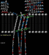
2D topology model of NadA predicted by TMBBPred server. Residues from 34-42, 89-97, 104-110 and 217-223 are transmembrane regions
Fig. 3
Fig. 3
Graphical representation of the most likely topology predicted by SPOCTOPUS. Network output: The two diagrams show the estimated preference for each residue to be located in different structural regions. The top diagram shows the preference of being either in (1) the hydrophobic part of the membrane, 0–13 A ° from the membrane center (M), (2) the membrane water-interface, 11–18 A ° from the membrane center (I), (3) a close loop region, 13–23 A ° from the membrane center (L), (4) a globular region, further than 23 A ° from the membrane (G). The bottom diagram shows the estimated preference of a particular residue to be located either on the inside (i) or on the outside (o) of the membrane. The raw data underlying these two plots can be found in the OCTOPUS network file
Fig. 4
Fig. 4
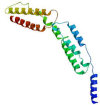
NadA 3D structure model predicted by SWISS MODEL
Fig. 5
Fig. 5
Model validations. Both global and local estimation of the quality of the obtained model are reasonable
Fig. 6
Fig. 6
Model evaluation. (a) Ramachandran plot of final NadA model. Number of residues in favored region: 177 (93.7%). Number of residues in allowed region: 9 (4.6%). Number of residues in outlier region: 3 (1.8%). (b)- Prosa protein structure anlysis results. Z score = -4.75. Overall quality of the ultimate model is acceptable
Fig. 7
Fig. 7
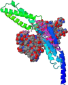
NadA ligand binding sites predicted by COFACTOR. NadA structure in contact with peptide ligand from lateral view. Ligand in the space filling model
Fig. 8
Fig. 8
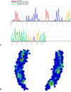
Functional and structural critical residues. (a) Colored illustration of conserved functional and structural residues predicted by conSeq server. The evolutionary rate of the sequence is obvious by color scale (see legend). Burial status of the location is labeled by b (buried) and e (exposed). Functionally or structurally importance of the residues were shown by f and s respectively. (b) Different direction of functional residues at the protein structure surface predicted by interproSurf
Fig. 9
Fig. 9
Linear B cell epitopes predicted by Bepipred server
Fig10
Fig10
B cell epitopes with the highest PI score. 2 linear (a) and 2 discontinuous (b) predicted epitopes are represented. Epitopes were mapped on 3D models using Discovery Studio Visualizer 2.5.5 software
Fig 11
Fig 11
GHECOM results for NadA Pocket detection.

Similar articles

Cited by

References

    1. Rouphael NG, Stephens DS. Neisseria meningitidis: biology, microbiology, and epidemiology. Methods Mol Biol. 2012;799:1–20. - PMC - PubMed
    1. Yazdankhah SP, Caugant DA. Neisseria meningitidis: an overview of the carriage state. J Med Microbiol. 2004;53:821–32. - PubMed
    1. Khater WS, Elabd SH. Identification of Common Bacterial Pathogens Causing Meningitis in Culture-Negative Cerebrospinal Fluid Samples Using Real-Time Polymerase Chain Reaction. Int J Microbiol. 2016;2016:5. - PMC - PubMed
    1. Gotschlich EC, Liu TY, Artenstein MS. Human immunity to the meningococcus. 3. Preparation and immunochemical properties of the group A, group B, and group C meningococcal polysaccharides. J Exp Med. 1969;129:1349–65. - PMC - PubMed
    1. Jafri RZ, Ali A, Messonnier NE, et al. Global epidemiology of invasive meningococcal disease. Popul Health Metr. 2013;11:17. - PMC - PubMed

LinkOut - more resources