. 2018 Sep 20;13(9):e0203657.
doi: 10.1371/journal.pone.0203657. eCollection 2018.

Propionate-producing bacteria in the intestine may associate with skewed responses of IL10-producing regulatory T cells in patients with relapsing polychondritis

Affiliations

Propionate-producing bacteria in the intestine may associate with skewed responses of IL10-producing regulatory T cells in patients with relapsing polychondritis

Jun Shimizu et al. PLoS One. .

Abstract

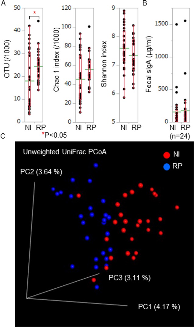
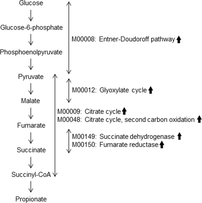
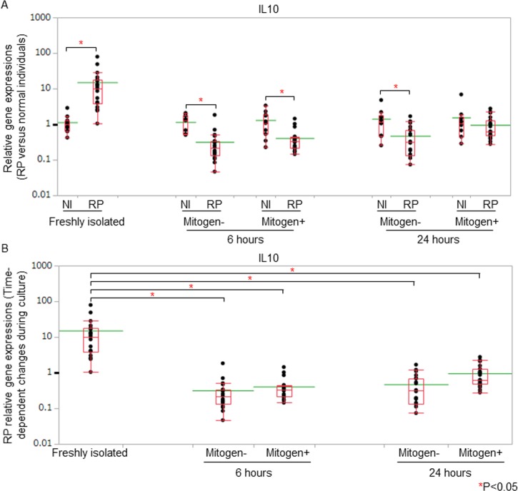
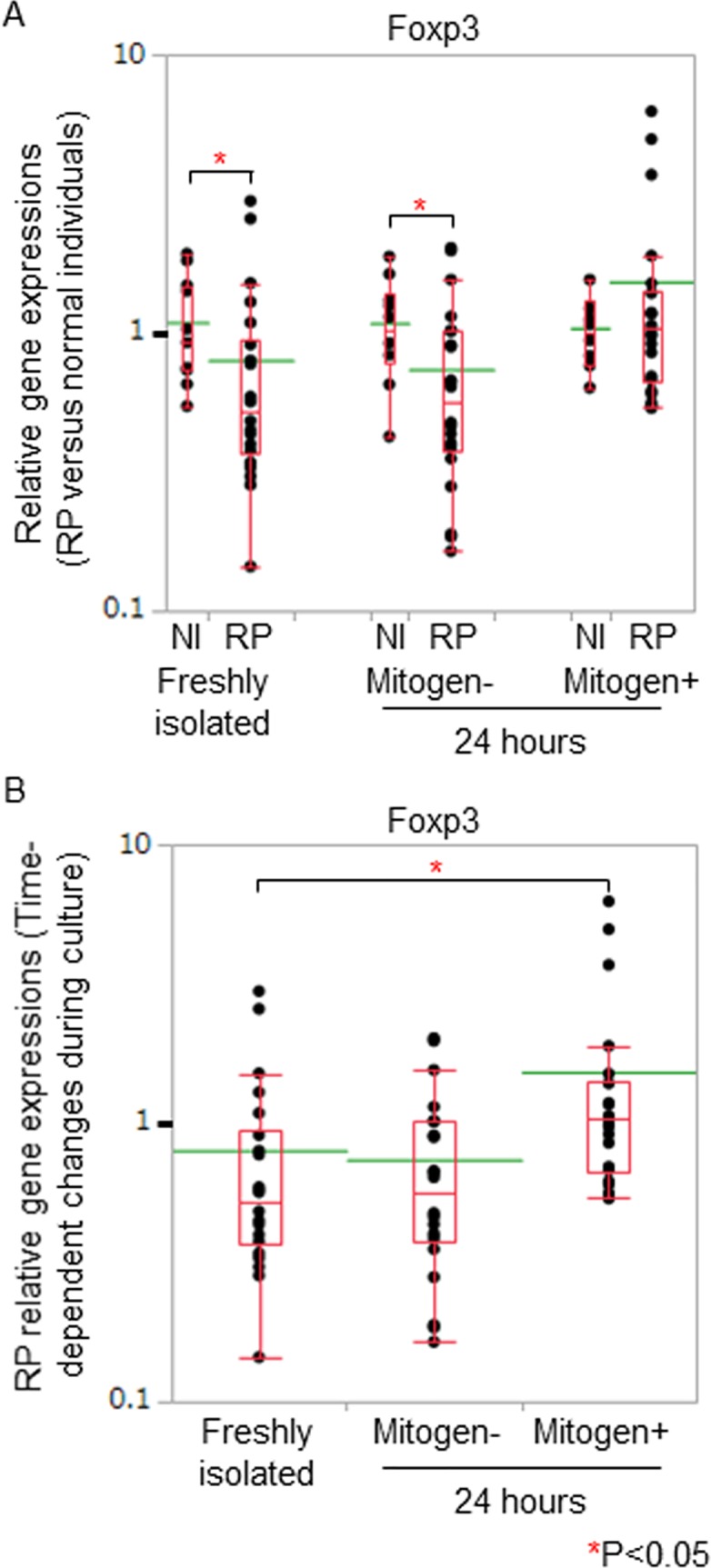
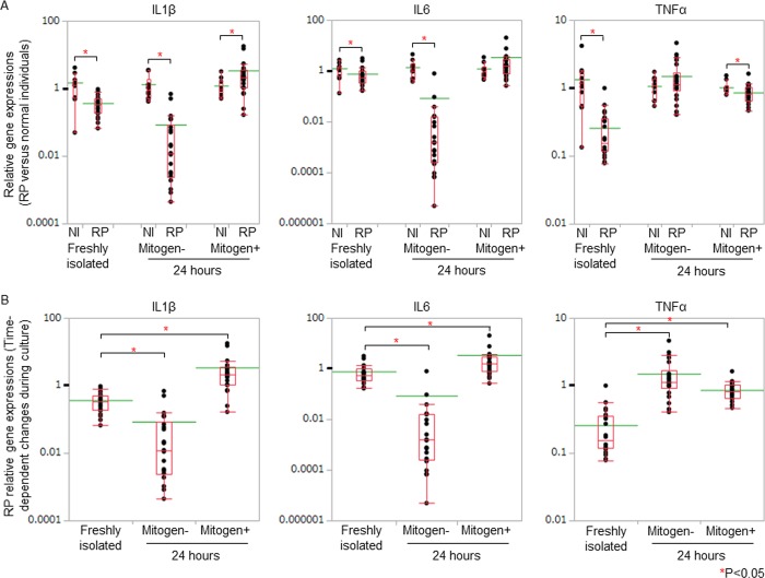
Relapsing polychondritis (RP) is an inflammatory disease of unknown causes, characterized by recurrent inflammation in cartilaginous tissues of the whole body. Recently, researchers have reported that, in mouse experiments, altered gut microbe-dependent T cell differentiation occurred in gut associated lymphoid tissues. Here, we investigated whether gut microbe alteration existed, and if so, the alteration affected peripheral T cell differentiation in patients with RP. In an analysis of gut microbiota, we found increased annotated species numbers in RP patients compared with normal individuals. In the RP gut microbiota, we observed several predominant species, namely Veillonella parvula, Bacteroides eggerthii, Bacteroides fragilis, Ruminococcus bromii, and Eubacterium dolichum, all species of which were reported to associate with propionate production in human intestine. Propionate is a short-chain fatty acid and is suggested to associate with interleukin (IL)10-producing regulatory T (Treg) cell differentiation in gut associated lymphoid tissues. IL10 gene expressions were moderately higher in freshly isolated peripheral blood mononuclear cells (PBMC) of RP patients than those of normal individuals. Six hours after the initiation of the cell culture, regardless of the presence and absence of mitogen stimulation, IL10 gene expressions were significantly lower in RP patients than those in normal individuals. It is well known that PBMC of patients with autoimmune and inflammatory diseases show hyporesponsiveness to mitogen stimulation. We suggest that, in RP patients, continuous stimulation of intestinal T cells by excessive propionate leads to the spontaneous IL10 production and a subsequent refractory period of T cells in patients with RP. The hyporesponsiveness of Treg cells upon activation may associate with inflammatory cytokine production of PBMC and subsequently relate to chondritis in RP patients.


Conflict of interest statement

The authors have declared that no competing interests exist.

Figures

Fig 1
Fig 1. Bacterial numbers and diversity of RP gut microbiota.
(A) We estimated OTU numbers (annotated species numbers) and α diversity scores (Chao 1 and Shannon indexes) of each sample. OTU numbers of RP patients (RP) were significantly higher than those of normal individuals (NI). Chao 1 and Shannon index scores were comparable between RP patients and normal individuals. (B) We measured fecal sIgA concentrations and compared the data between RP patients and normal individuals. Fecal sIgA concentrations were comparable between RP patients and normal individuals. These biological parameters of RP patients and normal individuals were displayed with dot plots. A box-plot and a mean level (green line) of each group of RP patients and normal individuals were indicated. (C) We estimated β diversity between RP patients and normal individuals. We showed the PCoA plots in a three dimensional structure where three axes and each contribution ratio were depicted. We exploratory evaluated the distance between the distribution of RP patients and that of normal individuals using two-sided Student's two-sample t-test and Monte Carlo permutations. We obtained a significant P value of the β diversity between RP patients and normal individuals in unweighted UniFrac PCoA (P = 0.01).
Fig 2
Fig 2. Schematic representation of succinate-propionate pathway and its alterations in patients with RP.
Several predominant gene functions of RP gut microbiota of this study (arrows) corresponded to several components of the succinate-propionate pathway. Veillonella species, Bacteroides species, and Acidaminococcus species utilize the pathway for the propionate production.
Fig 3
Fig 3. IL10 gene expressions on PBMC of RP patients.
(A) IL10 gene expressions of RP PBMC compared with those of normal PBMC. We measured IL10 gene expressions of freshly isolated, 6-hour-cultured, and 24-hour-cultured PBMC in RP patients (RP) and normal individuals (NI). Gene expressions of RP patients were estimated by the ΔΔCt method, followed by normalization of the titers relative to 2-ΔΔCt of normal individuals. The gene expression levels of normal individuals were defined as 1.000. IL10 gene expressions of freshly isolated PBMC were significantly higher in RP patients than those in normal individuals. IL10 gene expressions were significantly lower in RP patients than those of normal individuals 6 hours after the initiation of the cell culture, regardless of the presence and absence of mitogen stimulation. (B) Time-dependent changes of IL10 gene expressions of RP PBMC. We compared the gene expression data of the freshly isolated PBMC with those of cultured PBMC in patients with RP. IL10 gene expressions decreased significantly regardless of the presence and absence of mitogen stimulation in RP patients after the initiation of the cell culture. Relative gene expressions of RP patients against those of normal individuals were displayed with dot plots. A box-plot and a mean level (green line) of each group of RP patients and normal individuals were indicated.
Fig 4
Fig 4. Foxp3 gene expressions in PBMC of RP patients.
(A) We assessed RP relative gene expressions of Foxp3 by normalizing the titers to 2-ΔΔCt of normal individuals. The gene expression levels of normal individuals were defined as 1.000. Foxp3 gene expressions of RP freshly isolated PBMC were significantly lower than those of normal individuals. (B) We compared the gene expression data of the freshly isolated PBMC with those of cultured PBMC in patients with RP. Foxp3 gene expressions increased significantly in the presence of mitogen stimulation 24 hours after the initiation of the cell culture. Relative gene expressions of RP patients against those of normal individuals were displayed with dot plots. A box-plot and a mean level (green line) of each group of RP patients and normal individuals were indicated.
Fig 5
Fig 5. Inflammatory cytokine gene expressions in PBMC of RP patients.
(A) We assessed RP relative gene expressions of inflammatory cytokines by normalizing the titers to 2-ΔΔCt of normal individuals. The gene expression levels of normal individuals were defined as 1.000. IL1β gene expressions of RP patients were significantly higher than those of normal individuals in the presence of mitogen stimulation, 24 hours after the initiation of the cell culture. (B) We compared the gene expression data of the freshly isolated PBMC with those of cultured PBMC in patients with RP. After the initiation of the cell culture, in the absence of mitogen stimulation, TNFα gene expressions increased significantly in RP patients. With the stimulation, IL1β, IL6, and TNFα gene expressions increased significantly in RP patients. Relative gene expressions of RP patients against those of normal individuals were displayed with dot plots. A box-plot and a mean level (green line) of each group of RP patients and normal individuals were indicated.

References

    1. Letko E, Zafirakis P, Baltatzis S, Voudouri A, Livir-Rallatos C, Foster CS. Relapsing polychondritis: a clinical review. Semin Arthritis Rheum. 2002; 31: 384–395. - PubMed
    1. Oka H, Yamano Y, Shimizu J, Yudoh K, Suzuki N. A large-scale survey of patients with relapsing polychondritis in Japan. Inflamm Regen. 2014; 34: 149–156.
    1. Foidart JM, Abe S, Martin GR, Zizic TM, Barnett EV, Lawley TJ, et al. Antibodies to type II collagen in relapsing polychondritis. N Engl J Med. 1978; 299: 1203–1207. 10.1056/NEJM197811302992202 - DOI - PubMed
    1. Hansson AS, Heinegård D, Piette JC, Burkhardt H, Holmdahl R. The occurrence of autoantibodies to matrilin 1 reflects a tissue-specific response to cartilage of the respiratory tract in patients with relapsing polychondritis. Arthritis Rheum. 2001; 44: 2402–2412. - PubMed
    1. Sato T, Yamano Y, Tomaru U, Shimizu Y, Ando H, Okazaki T, et al. Serum level of soluble triggering receptor expressed on myeloid cells-1 as a biomarker of disease activity in relapsing polychondritis. Mod Rheumatol. 2014; 24: 129–136. 10.3109/14397595.2013.852854 - DOI - PubMed

Publication types

LinkOut - more resources