

A CARE-compliant article: Extranasal glial heterotopia in a female infant: A case report
- PMID: 30235657
- PMCID: PMC6160084
- DOI: 10.1097/MD.0000000000012000
A CARE-compliant article: Extranasal glial heterotopia in a female infant: A case report
Abstract
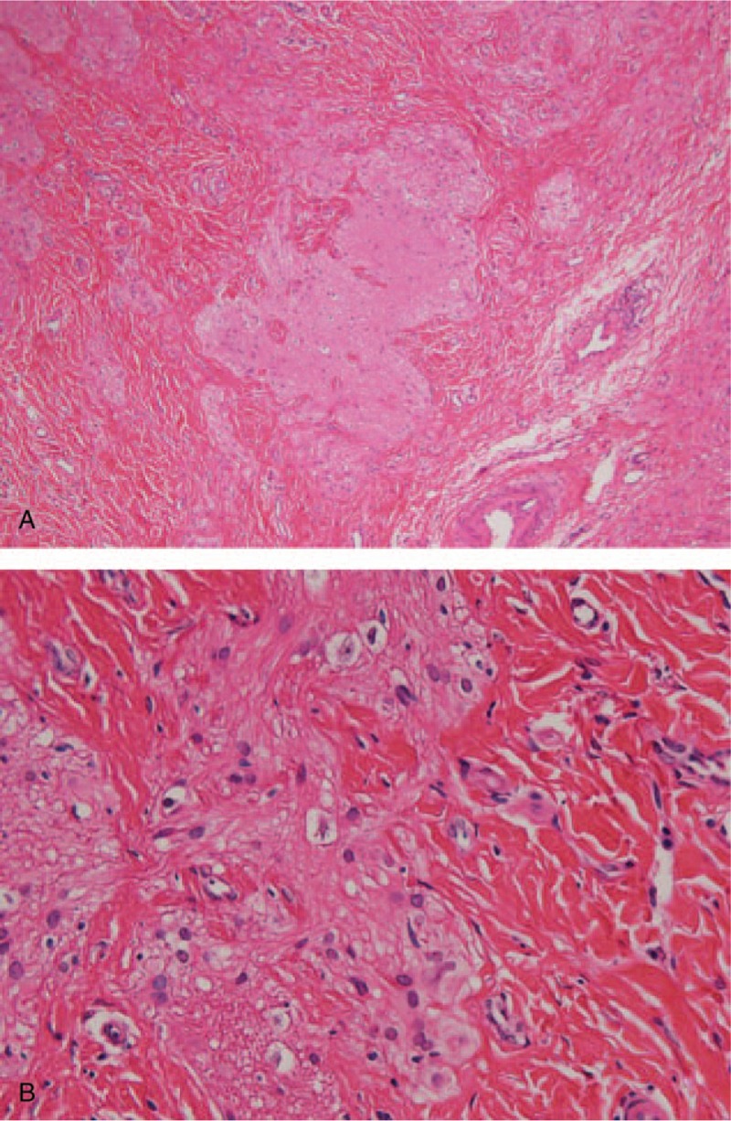
Rationale: Nasal glial heterotopia is a rare type of neoplasm consisting of meningothelial and/or neuroglial elements.
Patient concerns: A 17-month-old female infant was evaluated for treatment for a congenital mass present since birth on the right side of the nasal dorsum.
Diagnoses: The patient was preoperatively diagnosed with a congenital extranasal neoplasm.
Interventions: Surgery was performed under general anesthesia, and the mass was completely resected. The tissue was sent for histological examination, and the diagnosis was of extranasal glial heterotopia.
Outcomes: The surgical outcome was good, and no surgical site infection was recorded. After 6 months of follow-up, the girl was asymptomatic with no recurrence.
Lessons: Surgical excision, a curative method used to address extranasal glial heterotopia, resulted in no recurrence during the clinical follow-up period. The potential for an intracranial connection must always be kept in mind when considering how to surgically treat a congenital midline mass to prevent the risk of cerebrospinal fluid leakage.
Conflict of interest statement
The authors have no conflicts of interest to disclose.
Figures


References
-
- Rouev P, Dimov P, Shomov G. A case of nasal glioma in a new-born infant. Int J Pediatr Otorhinolaryngol 2001;58:91–4. - PubMed
-
- Hughes GB, Sharpino G, Hunt W, et al. Management of the congenital midline nasal mass: a review. Head Neck Surg 1980;2:222–33. - PubMed
-
- Bradley PJ, Singh SD. Congenital nasal masses: diagnosis and management. Clin Otolaryngol Allied Sci 1982;7:87–97. - PubMed
-
- Hedlund G. Congenital frontonasal masses: developmental anatomy, malformations, and MR imaging. Pediatr Radiol 2006;36:647–62. - PubMed
Publication types
MeSH terms
LinkOut - more resources
Full Text Sources
Other Literature Sources
Medical
