

Efficacy and adverse reactions of methotrexate in the treatment of ocular cicatricial pemphigoid: A case series study
- PMID: 30235689
- PMCID: PMC6160102
- DOI: 10.1097/MD.0000000000012338
Efficacy and adverse reactions of methotrexate in the treatment of ocular cicatricial pemphigoid: A case series study
Abstract
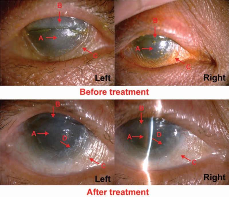
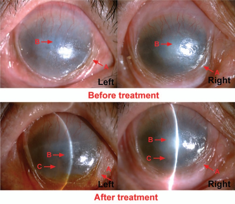
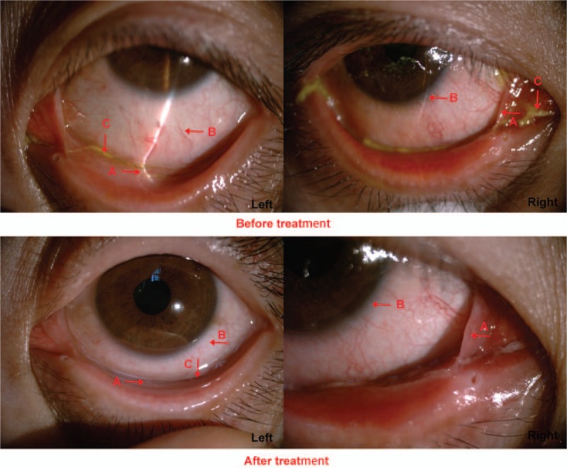
The application of methotrexate (MTX) in the treatment of autoimmune diseases has been gradually increasing, but reports of MTX treatment for advanced ocular cicatricial pemphigoid (OCP) are extremely rare. This study investigated the efficacy and adverse reactions of low-dose MTX in patients with OCP.This was a retrospective, noncontrolled, case series study. Eleven patients diagnosed with advanced OCP (4 cases in stage III and 7 cases in stage IV) were enrolled. Treatment by oral administration of MTX (7.5 ± 2.5 mg) alone was performed. Visual acuity of the patients, conjunctival inflammation, cicatrization, ocular surface keratinization, and toxic side effects of drugs were evaluated.All patients enrolled in this study were females aged 32 to 83 years. Patients were followed up for 4 to 33 months. Low-dose MTX improved visual acuity of 3 cases (6 eyes, 27.3%). Conjunctival inflammation of 5 patients (10 eyes) rested after treatment, and conjunctival inflammation of 3 cases (6 eyes, 27.3%) was decreased with an effective rate of 72.7%. Cicatrices of 8 cases (15 eyes) showed degeneration after treatment with an effective rate of 71.4% (15/21). Ocular surface keratinization receded in 4 cases with an effective rate of 66.7%. None of the patients discontinued the treatment due to severe toxic side effects. All patients tolerated mild drug-induced gastrointestinal reactions. Three patients terminated the treatment in advance after 4 to 6 months due to no improvement in the disease condition.Observation of clinical efficacy and safety findings demonstrated that low-dose MTX can be used to treat patients with advanced OCP.
Conflict of interest statement
The authors have no conflicts of interest to disclose.
Figures

Similar articles
-
Methotrexate therapy for ocular cicatricial pemphigoid.Ophthalmology. 2004 Apr;111(4):796-801. doi: 10.1016/j.ophtha.2003.07.010. Ophthalmology. 2004. PMID: 15051215
-
The effect of treatment and its related side effects in patients with severe ocular cicatricial pemphigoid.Ophthalmology. 2002 Jan;109(1):111-8. doi: 10.1016/s0161-6420(01)00863-6. Ophthalmology. 2002. PMID: 11772589
-
[Ocular Cicatricial Pemphigoid - a Retrospective Study].Cesk Slov Oftalmol. 2016 Feb;72(1):283-92. Cesk Slov Oftalmol. 2016. PMID: 27041284 Czech.
-
Ocular cicatricial pemphigoid: manifestations and management.Curr Allergy Asthma Rep. 2005 Jul;5(4):333-8. doi: 10.1007/s11882-005-0078-9. Curr Allergy Asthma Rep. 2005. PMID: 15967079 Review.
-
[Diagnostics and pharmacological treatment of ocular cicatrical pemphigoid].Klin Oczna. 2005;107(10-12):725-7. Klin Oczna. 2005. PMID: 16619831 Review. Polish.
Cited by
-
Ocular Cicatricial Pemphigoid, Sjögren's Syndrome, and Hashimoto's Thyroiditis as a Multiple Autoimmune Syndrome: A case report.Eur J Ophthalmol. 2022 Jul;32(4):NP52-NP55. doi: 10.1177/1120672121996637. Epub 2021 Feb 18. Eur J Ophthalmol. 2022. PMID: 33601905 Free PMC article.
-
Smart Eye Camera: A Validation Study for Evaluating the Tear Film Breakup Time in Human Subjects.Transl Vis Sci Technol. 2021 Apr 1;10(4):28. doi: 10.1167/tvst.10.4.28. Transl Vis Sci Technol. 2021. PMID: 34004005 Free PMC article.
-
Ocular mucous membrane pemphigoid: a review.Immunol Res. 2019 Jun;67(2-3):280-289. doi: 10.1007/s12026-019-09087-7. Immunol Res. 2019. PMID: 31327149 Review.
-
European Guidelines (S3) on diagnosis and management of mucous membrane pemphigoid, initiated by the European Academy of Dermatology and Venereology - Part II.J Eur Acad Dermatol Venereol. 2021 Oct;35(10):1926-1948. doi: 10.1111/jdv.17395. Epub 2021 Jul 26. J Eur Acad Dermatol Venereol. 2021. PMID: 34309078 Free PMC article.
References
-
- Sobolewska B, Deuter C, Zierhut M. Current medical treatment of ocular mucous membrane pemphigoid. Ocul Surf 2013;11:259–66. - PubMed
-
- Levine MR. Black EH, Nesi FA, Calvano CJ, Gladstone GJ, Levine MR. Ocular Cicatricial Pemphigoid. Smith and Nesi's Ophthalmic Plastic and Reconstructive Surgery. New York, NY: Springer New York; 2012. 355–7.
-
- Chan LS, Ahmed AR, Anhalt GJ, et al. The first international consensus on mucous membrane pemphigoid: definition, diagnostic criteria, pathogenic factors, medical treatment, and prognostic indicators. Arch Dermatol 2002;138:370–9. - PubMed
-
- Saw VP, Dart JK. Ocular mucous membrane pemphigoid: diagnosis and management strategies. Ocul Surf 2008;6:128–42. - PubMed
Publication types
MeSH terms
Substances
LinkOut - more resources
Full Text Sources
Other Literature Sources
Medical
