

Comparison of 1.5- and 3.0-T magnetic resonance imaging for evaluating lesions of the knee: A systematic review and meta-analysis (PRISMA-compliant article)
- PMID: 30235710
- PMCID: PMC6160024
- DOI: 10.1097/MD.0000000000012401
Comparison of 1.5- and 3.0-T magnetic resonance imaging for evaluating lesions of the knee: A systematic review and meta-analysis (PRISMA-compliant article)
Abstract
Background: With conflicting results in the literature, it remains unclear whether a higher field strength automatically increases the sensitivity and specificity of magnetic resonance imaging (MRI) for detecting pathological lesions in the knee. Therefore, we performed a systematic review and meta-analysis of studies comparing the diagnostic accuracy of 1.5- and 3.0-T MRI for lesions within the knee.
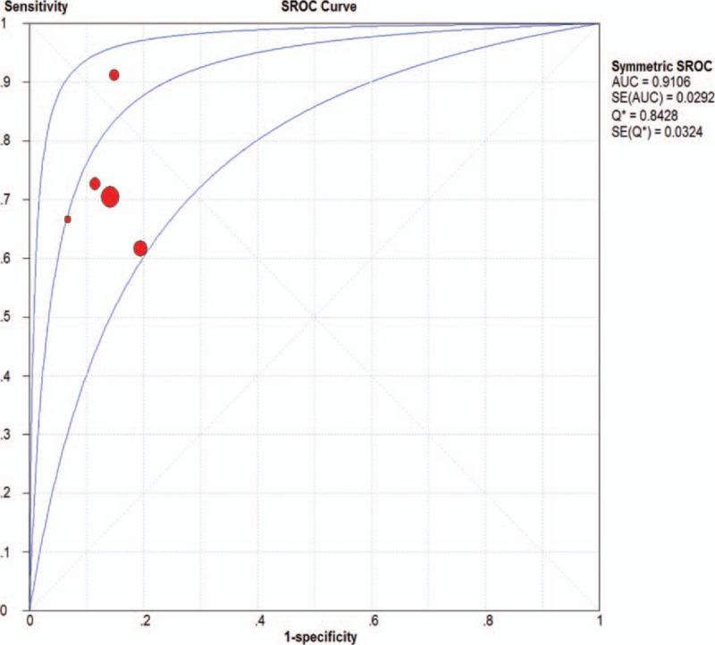
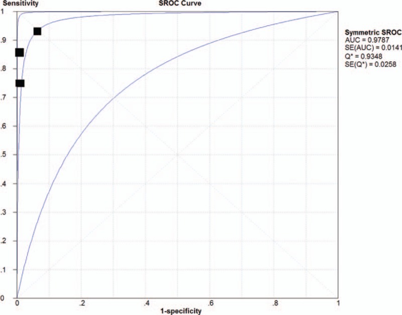
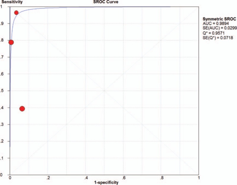
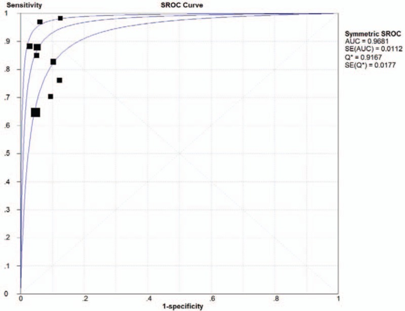
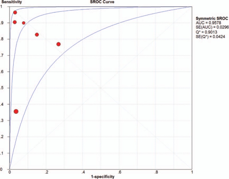
Methods: Sixteen studies were included in the meta-analysis of the diagnostic accuracy of MRI for lesions of the knee joint, and areas under the curve (AUC) derived from the summary receiver operating characteristic curve analysis were determined for comparison of the diagnostic accuracy with differing magnetic field strength as well as for lesions in different tissues of the knee. Separate meta-analyses were performed for the diagnosis of lesions within articular cartilage, ligaments, and meniscus.
Results: For lesions within the articular cartilage, the AUC for 1.5-T MRI differed significantly from that for 3.0-T MRI (Z = 3.4, P < .05). However, for lesions within the ligaments and meniscus, the AUC values for 1.5-T MRI did not differ significantly from those for 3.0-T MRI (Z = 0.32, P > .05, and Z = 0.33, P > .05, respectively).
Conclusion: Our results indicate that both 1.5-T and 3.0-T MRI offer high diagnostic accuracy and clinical relevance for knee injuries involving the meniscus or a ligament. However, the present meta-analysis indicates that 3.0-T MRI does offer greater diagnostic accuracy than 1.5-T MRI for articular cartilage lesions.
Conflict of interest statement
The authors have no conflicts of interest to disclose.
Figures

Similar articles
-
Magnetic resonance perfusion for differentiating low-grade from high-grade gliomas at first presentation.Cochrane Database Syst Rev. 2018 Jan 22;1(1):CD011551. doi: 10.1002/14651858.CD011551.pub2. Cochrane Database Syst Rev. 2018. PMID: 29357120 Free PMC article.
-
A systematic review of duplex ultrasound, magnetic resonance angiography and computed tomography angiography for the diagnosis and assessment of symptomatic, lower limb peripheral arterial disease.Health Technol Assess. 2007 May;11(20):iii-iv, xi-xiii, 1-184. doi: 10.3310/hta11200. Health Technol Assess. 2007. PMID: 17462170
-
Imaging modalities for the detection of posterior pelvic floor disorders in women with obstructed defaecation syndrome.Cochrane Database Syst Rev. 2021 Sep 23;9(9):CD011482. doi: 10.1002/14651858.CD011482.pub2. Cochrane Database Syst Rev. 2021. PMID: 34553773 Free PMC article.
-
Magnetic resonance imaging for the diagnosis of hepatocellular carcinoma in adults with chronic liver disease.Cochrane Database Syst Rev. 2022 May 6;5(5):CD014798. doi: 10.1002/14651858.CD014798.pub2. Cochrane Database Syst Rev. 2022. PMID: 35521901 Free PMC article.
-
Magnetic resonance imaging is able to detect patellofemoral focal cartilage injuries: a systematic review with meta-analysis.Knee Surg Sports Traumatol Arthrosc. 2023 Jun;31(6):2469-2481. doi: 10.1007/s00167-022-07203-z. Epub 2022 Oct 20. Knee Surg Sports Traumatol Arthrosc. 2023. PMID: 36266368
Cited by
-
Patterns of Articular Cartilage Thickness in Pediatric and Adolescent Knees: A Magnetic Resonance Imaging-Based Study.Arthrosc Sports Med Rehabil. 2021 Feb 2;3(2):e381-e390. doi: 10.1016/j.asmr.2020.09.029. eCollection 2021 Apr. Arthrosc Sports Med Rehabil. 2021. PMID: 34027446 Free PMC article.
-
MRI Advancements in Musculoskeletal Clinical and Research Practice.Radiology. 2023 Aug;308(2):e230531. doi: 10.1148/radiol.230531. Radiology. 2023. PMID: 37581501 Free PMC article. Review.
-
ESR essentials: MRI of the knee-practice recommendations by ESSR.Eur Radiol. 2024 Oct;34(10):6590-6599. doi: 10.1007/s00330-024-10706-7. Epub 2024 Mar 27. Eur Radiol. 2024. PMID: 38536461 Free PMC article. Review.
-
Meniscal Extrusion: Diagnosis, Etiology, and Treatment Options.Curr Rev Musculoskelet Med. 2023 Jul;16(7):316-327. doi: 10.1007/s12178-023-09840-4. Epub 2023 May 16. Curr Rev Musculoskelet Med. 2023. PMID: 37191818 Free PMC article. Review.
-
Evaluation of Osteochondral Allograft Transplant Using In-Office Needle Arthroscopy.Arthrosc Tech. 2022 Nov 17;11(12):e2243-e2248. doi: 10.1016/j.eats.2022.08.032. eCollection 2022 Dec. Arthrosc Tech. 2022. PMID: 36632378 Free PMC article.
References
-
- Kijowski R, Blankenbaker DG, Davis KW, et al. Comparison of 1.5- and 3.0-T MR imaging for evaluating the articular cartilage of the knee joint. Radiology 2009;250:839–48. - PubMed
-
- Laprade RF, Ho CP, James E, et al. Diagnostic accuracy of 3.0 T magnetic resonance imaging for the detection of meniscus posterior root pathology. Knee Surg Sports Traumatol Arthrosc 2015;23:152–7. - PubMed
-
- Phelan N, Rowland P, Galvin R, et al. A systematic review and meta-analysis of the diagnostic accuracy of MRI for suspected ACL and meniscal tears of the knee. Knee Surg Sports Traumatol Arthrosc 2016;24:1525–39. - PubMed
-
- Figueroa D, Calvo R, Vaisman A, et al. Knee chondral lesions: incidence and correlation between arthroscopic and magnetic resonance findings. Arthroscopy 2007;23:312–5. - PubMed
-
- Vaz CE, Camargo OP, Santana PJ, et al. Accuracy of magnetic resonance in identifying traumatic intraarticular knee lesions. Clinics (Sao Paulo) 2005;60:445–50. - PubMed
Publication types
MeSH terms
LinkOut - more resources
Full Text Sources
Other Literature Sources
Medical
