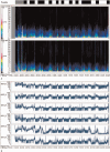
Detection of seizure patterns with multichannel amplitude-integrated EEG and the color density spectral array in the adult neurology intensive care unit
- PMID: 30235767
- PMCID: PMC6160116
- DOI: 10.1097/MD.0000000000012514
Detection of seizure patterns with multichannel amplitude-integrated EEG and the color density spectral array in the adult neurology intensive care unit
Abstract
This study's purpose was to determine the sensitivity, false-positive and false-negative of seizure detection in adult intensive care by amplitude-integrated electroencephalography (aEEG) and color density spectral array (CDSA).30 continuous electroencephalogram (EEG) recordings were randomly performed in 3 digital EEG-recording machines, 3 specialized neurophysiologists participated in this study, underwent 4 hours of training of CDSA and aEEG, marked any epochs suspected to be seizures without access to the raw EEG. The results will be compared and analyzed with continuous EEG reading to consider sensitivity, positive or negative rate.The recordings in this study, comprised 720 hours of EEG containing a total of 435 seizures. The median sensitivity for seizure identification was 80% of CDSA and 81.3% of aEEG, Median false-positive was 4 per 24 hours of CDSA, and 2 per 24 hours of aEEG display, Median false-negative was 4 per 24 hours of CDSA, and 4 per 24 hours of aEEG display. The time spent in identification of seizures by CDSA and aEEG was much time-saving than continuous EEG-reading.In this study, both CDSA and aEEG have a higher sensitivity but lower false-positive or missed rate in the interpretation of seizure identification in adult NICU.
Conflict of interest statement
The authors have no conflicts of interest to disclose.
Figures

Similar articles
-
Seizure identification in the ICU using quantitative EEG displays.Neurology. 2010 Oct 26;75(17):1501-8. doi: 10.1212/WNL.0b013e3181f9619e. Epub 2010 Sep 22. Neurology. 2010. PMID: 20861452 Free PMC article.
-
Seizure Detection by Critical Care Providers Using Amplitude-Integrated Electroencephalography and Color Density Spectral Array in Pediatric Cardiac Arrest Patients.Pediatr Crit Care Med. 2017 Apr;18(4):363-369. doi: 10.1097/PCC.0000000000001099. Pediatr Crit Care Med. 2017. PMID: 28234810 Free PMC article. Clinical Trial.
-
Non-expert use of quantitative EEG displays for seizure identification in the adult neuro-intensive care unit.Epilepsy Res. 2015 Jan;109:48-56. doi: 10.1016/j.eplepsyres.2014.10.013. Epub 2014 Oct 28. Epilepsy Res. 2015. PMID: 25524842
-
Amplitude-integrated electroencephalography for seizure detection in newborn infants.Semin Fetal Neonatal Med. 2018 Jun;23(3):175-182. doi: 10.1016/j.siny.2018.02.003. Epub 2018 Feb 13. Semin Fetal Neonatal Med. 2018. PMID: 29472139 Review.
-
The role of electroencephalogram in neonatal seizure detection.Expert Rev Neurother. 2018 Feb;18(2):95-100. doi: 10.1080/14737175.2018.1413352. Epub 2017 Dec 8. Expert Rev Neurother. 2018. PMID: 29199490 Review.
Cited by
-
Seizure burden in preterm infants and smaller brain volume at term-equivalent age.Pediatr Res. 2022 Mar;91(4):955-961. doi: 10.1038/s41390-021-01542-2. Epub 2021 Apr 26. Pediatr Res. 2022. PMID: 33903729 Free PMC article.
-
Comparative analysis of background EEG activity based on MRI findings in neonatal hypoxic-ischemic encephalopathy: a standardized, low-resolution, brain electromagnetic tomography (sLORETA) study.BMC Neurol. 2022 Jun 3;22(1):204. doi: 10.1186/s12883-022-02736-9. BMC Neurol. 2022. PMID: 35659637 Free PMC article.
-
Quantitative EEG-Based Seizure Estimation in Super-Refractory Status Epilepticus.Neurocrit Care. 2022 Jun;36(3):897-904. doi: 10.1007/s12028-021-01395-x. Epub 2021 Nov 17. Neurocrit Care. 2022. PMID: 34791594 Free PMC article.
-
Quantitative Continuous EEG: Bridging the Gap Between the ICU Bedside and the EEG Interpreter.Neurocrit Care. 2019 Jun;30(3):499-504. doi: 10.1007/s12028-019-00694-8. Neurocrit Care. 2019. PMID: 30788706 No abstract available.
-
Recent applications of quantitative electroencephalography in adult intensive care units: a comprehensive review.J Neurol. 2022 Dec;269(12):6290-6309. doi: 10.1007/s00415-022-11337-y. Epub 2022 Aug 19. J Neurol. 2022. PMID: 35986096 Free PMC article. Review.
References
-
- Friedman D, Claassen J, Hirsch LJ. Continuous electroencephalogram monitoring in the intensive care unit. Anesth Analg 2009;109:506–23. - PubMed
-
- DeLorenzo RJ, Waterhouse EJ, Towne AR, et al. Persistent nonconvulsive status epilepticus after the control of convulsive status epilepticus. Epilepsia 1998;39:833–40. - PubMed
-
- Scher MS, Painter MJ, Bergman I, et al. EEG diagnosis of neonatal seizures: clinical correlations and outcome. Pediatr Neurol 1989;5:17–24. - PubMed
-
- Clancy RR, Legido ADL. Occult neonatal seizures. Epilepsia 1998;29:256–61. - PubMed
-
- Weiner SP, Painter MJ, Geva D, et al. Neonatal seizures: electroclinical dissociation. Pediatr Neurol 1991;7:363–8. - PubMed
Publication types
MeSH terms
LinkOut - more resources
Full Text Sources
Other Literature Sources
Medical

