. 2018 Sep 20;11(1):521.
doi: 10.1186/s13071-018-3092-1.

The marsupial trypanosome Trypanosoma copemani is not an obligate intracellular parasite, although it adversely affects cell health

Affiliations

The marsupial trypanosome Trypanosoma copemani is not an obligate intracellular parasite, although it adversely affects cell health

Crystal Cooper et al. Parasit Vectors. .

Abstract

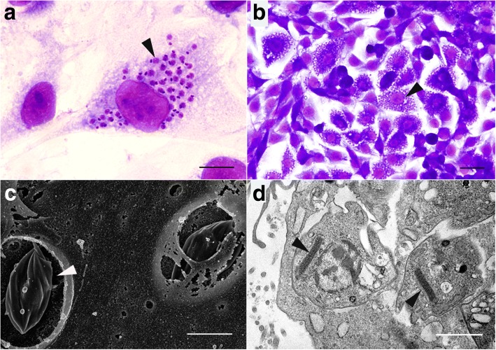
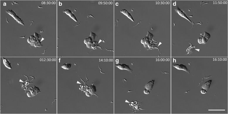
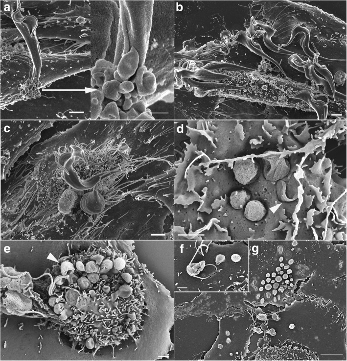
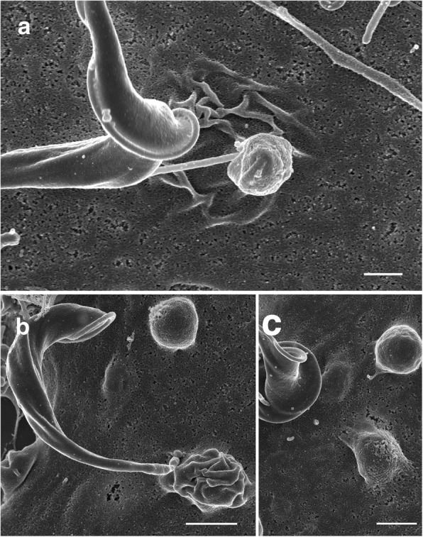
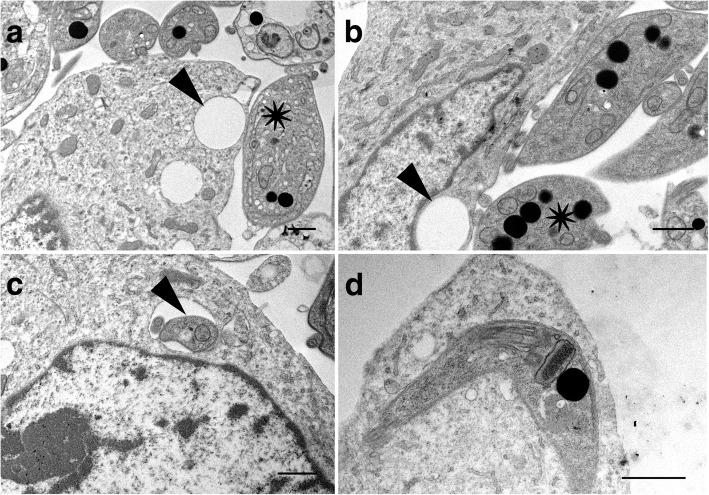
Background: Trypanosoma cruzi invades and replicates inside mammalian cells, which can lead to chronic Chagas disease in humans. Trypanosoma copemani infects Australian marsupials and recent investigations indicate it may be able to invade mammalian cells in vitro, similar to T. cruzi. Here, T. cruzi 10R26 strain (TcIIa) and two strains of T. copemani [genotype 1 (G1) and genotype 2 (G2)] were incubated with marsupial cells in vitro. Live-cell time-lapse and fluorescent microscopy, combined with high-resolution microscopy (transmission and scanning electron microscopy) were used to investigate surface interactions between parasites and mammalian cells.

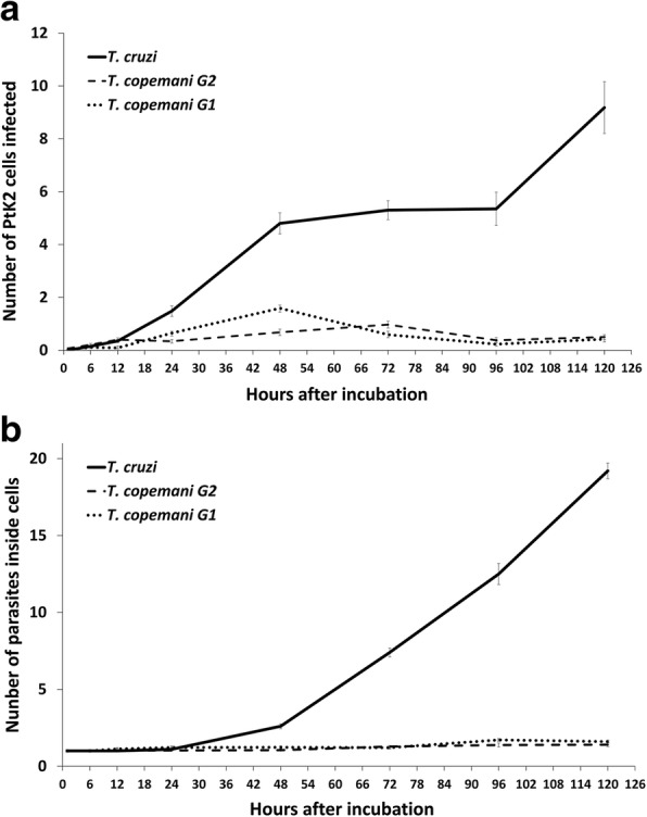
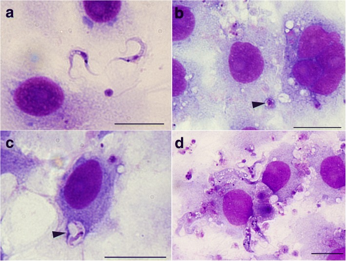
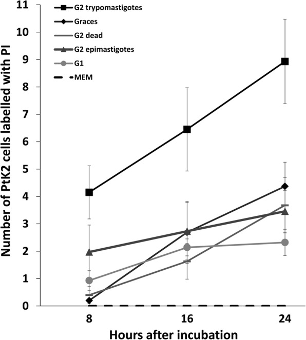
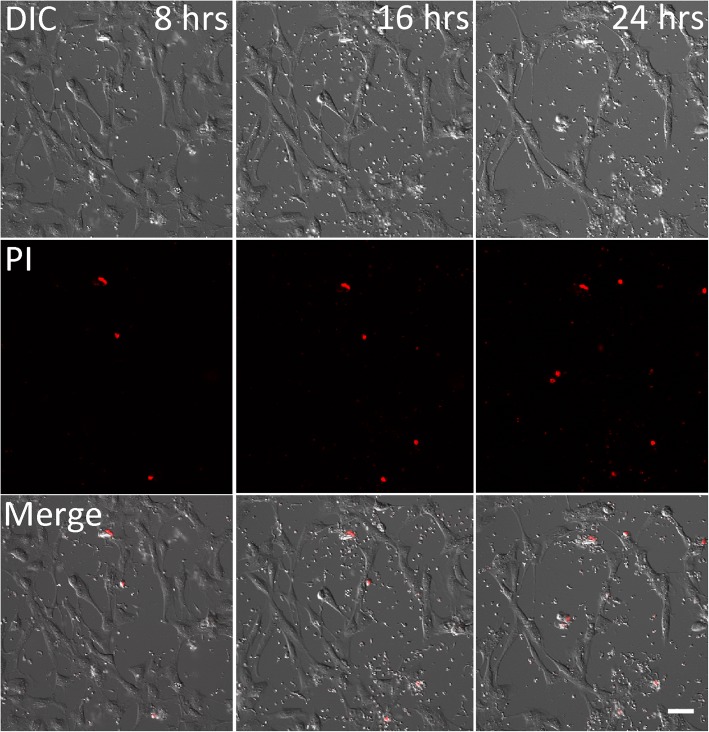
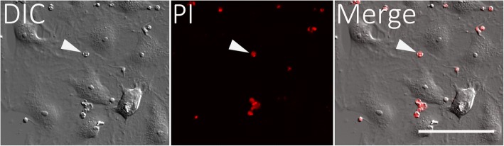
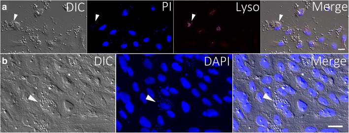
Results: The number of parasites invading cells was significantly higher in T. cruzi compared to either genotype of T. copemani, between which there was no significant difference. While capable of cellular invasion, T. copemani did not multiply in host cells in vitro as there was no increase in intracellular amastigotes over time and no release of new trypomastigotes from host cells, as observed in T. cruzi. Exposure of host cells to G2 trypomastigotes resulted in increased host cell membrane permeability within 24 h of infection, and host cell death/blebbing was also observed. G2 parasites also became embedded in the host cell membrane.

Conclusions: Trypanosoma copemani is unlikely to have an obligate intracellular life-cycle like T. cruzi. However, T. copemani adversely affects cell health in vitro and should be investigated in vivo in infected host tissues to better understand this host-parasite relationship. Future research should focus on increasing understanding of the T. copemani life history and the genetic, physiological and ecological differences between different genotypes.

Keywords: Australia; Host-parasite interactions; Marsupials; Trypanosoma copemani; Trypanosoma cruzi.


Conflict of interest statement

Ethics approval and consent to participate

Not applicable. No animal tissues or human samples were used in this study and ethics approval was not required.

Consent for publication

Not applicable.

Competing interests

The authors declare that they have no competing interests.

Publisher’s Note

Springer Nature remains neutral with regard to jurisdictional claims in published maps and institutional affiliations.

Figures

Fig. 1
Fig. 1
Trypanosoma cruzi and T. copemani G1 and G2 infecting potoroo kidney epithelial (PtK2) cells over 5 days. a Number of cells from 9000 (300 cells from 10 culture wells in 3 repeated experiments) (mean ± SE) infected with parasites over the five days. b Number of intracellular parasites inside 9000 cells (300 cells from 10 culture wells in 3 repeated experiments) (mean ± SE) in infected cells over five days
Fig. 2
Fig. 2
Microscopy of Trypanosoma cruzi infecting potoroo kidney epithelial (PtK2) cells. a Light micrograph of Diff-Quik stained cells and parasites after 96 h incubation showing a single cell with amastigotes inside, as recognisable by their round nucleus and disc shaped kinetoplast (arrowhead). b Heavily infected area of PtK2 cells (arrowhead) following incubation with T. cruzi for 120 h and stained with Diff-Quik. c Scanning electron micrograph of T. cruzi infecting PtK2 cells. The dying cell membranes were broken fortuitously exposing developing T. cruzi amastigotes (arrowhead) inside. d Transmission electron micrograph of T. cruzi amastigotes developing inside PtK2 cells. T. cruzi is recognisable by the dense elongate kinetoplasts (arrowheads). Scale-bars: a, 10 μm; b, 20 μm; c, 2 μm; d, 1 μm
Fig. 3
Fig. 3
Microscopy of Trypanosoma copemani G2 incubated with potoroo kidney epithelial (PtK2) cells. a Light microscopy of G2 trypomastigotes. b Light microscopy of G2 amastigote-like cells inside PtK2 cells (arrowhead). c G2 trypomastigote inside a vacuole within the cell (arrowhead). d G2 attached to the outside of cells. All images are stained with Diff-Quik. Scale-bars: 20 μm
Fig. 4
Fig. 4
The number (mean ± SE) of potoroo kidney epithelial (PtK2) cells displaying propidium iodine (PI) fluorescence, and thus with permeabilised or compromised membranes, after incubation with Trypanosoma copemani for 24 h. T. copemani G2 trypomastigotes, G2 epimastigotes and G1 trypomastigotes were incubated with PtK2 cells in MEM media for 24 h. Controls included dead parasites (G2 trypomastigotes), PtK2 cells in MEM without parasites and PtK2 cells in Grace’s media without FCS or parasites. Cells in 3 separate fields of view were counted (20×) and experiments were repeated twice to gain the proportion of cells displaying PI fluorescence for each treatment
Fig. 5
Fig. 5
Trypanosoma copemani G2 and potoroo epithelial kidney (PtK2) cells incubated with propidium iodide (PI). Cell uptake of PI increases over time when exposed to G2 trypomastigotes: 8 h; 16 h; 24 h. Channels are split into differential interference contrast (DIC), PI, and both channels merged (Merge). See Additional file 1: Figure S1 and Additional file 4 for higher magnification figures and a live-cell time-lapse video. Scale-bar: 50 μm
Fig. 6
Fig. 6
Multiple attachment of Trypanosoma copemani G2 trypomastigotes to Vero cells at different time-points during an 8 h incubation, 8 h after parasites were added to cells. The video shows parasite attachment can cause cell detachment from the glass substrate (indicated by arrowheads). Each panel a-h shows time after incubation with G2. See Additional file 6 for the live-cell time-lapse video. Scale-bars: 50 μm
Fig. 7
Fig. 7
High resolution scanning electron micrographs of Trypanosoma copemani G2 trypomastigotes after incubation with Vero cells for 24 (a-c) and 48 h (d-g). a Parasite attached to cell that is blebbing around the cell attachment site and an arrow showing higher magnification of the same site. b Attachment of multiple parasites to a cell that appears to be blebbing. c Attachment of multiple parasites to a cell that appears to be blebbing. d Parasites embedded in the cell membrane. Arrow points to a visible flagellum, e Parasites embedded in the cell surface and some retain long flagella that extend out of the cell (arrow). f Parasite that appears to be degrading with a flagellum on the surface of a cell g Multiple parasites attached to the cell surface that appear to be degrading. Scale-bars: a, 2 μm and 1 μm; b-f, 2 μm; g 10 μm
Fig. 8
Fig. 8
High resolution scanning electron micrographs of Trypanosoma copemani G2 after incubation with potoroo epithelial kidney (PtK2) cells for 24 h. a Attached T. copemani trypomastigotes and external amastigote attached to the cell surface. b Attached T. copemani trypomastigote. c Blebbing in PtK2 cell. Scale-bars: 1 μm
Fig. 9
Fig. 9
Trypanosoma copemani G1 after 24 h incubation with potoroo kidney epithelial cells and propidium iodide (PI). Amastigote-like cells are recognisable by the round appearance and the presence of PI indicates their membranes are compromised (arrow). Channels are split into DIC, PI, and both channels merged. Scale-bar: 50 μm
Fig. 10
Fig. 10
Transmission electron micrographs of potoroo kidney epithelial cells (PtK2) after incubation with Trypanosoma copemani G2 for 48 h. a Parasite attached to the surface of a cell. Parasite indicated by an asterisk is attached to the surface of the cell with a vacuole (arrow) underneath. b Two parasites attached to the surface of a cell one parasite (asterisk) with a vacuole underneath (arrow). c T. copemani inside a cell, inside a vacuole recognisable by the flagellum (arrow). d T. copemani attached to a cell, recognisable by the kinetoplast (arrow). Scale-bars: 1 μm
Fig. 11
Fig. 11
a Trypanosoma copemani infecting potoroo kidney epithelial (PtK2) cells in vitro after 24 h (fixed samples). Parasites and cells were stained with DAPI and Lysotracker®. DIC, DAPI, and Lysotracker® channels are split to show individual staining, and the merged panel is shown last. Arrows point to a cell with attached parasites and lysosomes that are not localised in the same place as parasite attachment. Cells are recognisable by their larger nuclei and parasites by their smaller nuclei stained with DAPI. b Trypanosoma cruzi infecting potoroo kidney epithelial (PtK2) cells in vitro after 72 h (fixed samples). Arrow indicates an infected cell. Parasites are recognisable by their smaller nucleus and kinetoplast stained with DAPI. Cells are distinguishable by their larger DAPI stained nuclei compared to the smaller parasite nuclei. Channels are split into DIC, DAPI and both channels merged. Scale-bar: 20 μm

Similar articles

Cited by

References

    1. Clayton J. Chagas disease 101. Nature. 2010;465:S4–S5. doi: 10.1038/nature09220. - DOI - PubMed
    1. WHO. Chagas disease (American trypanosomiasis). World Health Organ Fact Sheet 340. 2015. http://www.who.int/mediacentre/factsheets/fs340/en/. Accessed 28 Jul 2018.
    1. Schmunis GA, Yadon ZE. Chagas disease: a Latin American health problem becoming a world health problem. Acta Trop. 2006;115:14–21. doi: 10.1016/j.actatropica.2009.11.003. - DOI - PubMed
    1. Munoz-Saravia SG, Haberland A, Wallukat G, Schimke I. Chronic Chagas’ heart disease: a disease on its way to becoming a worldwide health problem: epidemiology, etiopathology, treatment, pathogenesis and laboratory medicine. Heart Fail Rev. 2010;17:45–64. doi: 10.1007/s10741-010-9211-5. - DOI - PubMed
    1. Salazar R, Castillo-neyra R, Tustin AW, Borrini-mayorı K, Naquira C, Levy MZ. Bed bugs (Cimex lectularius) as vectors of Trypanosoma cruzi. Am J Trop Med Hyg. 2015;92:331–335. doi: 10.4269/ajtmh.14-0483. - DOI - PMC - PubMed

MeSH terms

LinkOut - more resources