Comparative Study
. 2018 Dec;188(12):2717-2728.
doi: 10.1016/j.ajpath.2018.08.010. Epub 2018 Sep 18.

A Contrast in Pathogenic Responses between C57BL/6J and BALB/cJ Mice Using a Model of Retinal Injury

Affiliations
Comparative Study

A Contrast in Pathogenic Responses between C57BL/6J and BALB/cJ Mice Using a Model of Retinal Injury

Haoshen Shi et al. Am J Pathol. 2018 Dec.

Abstract

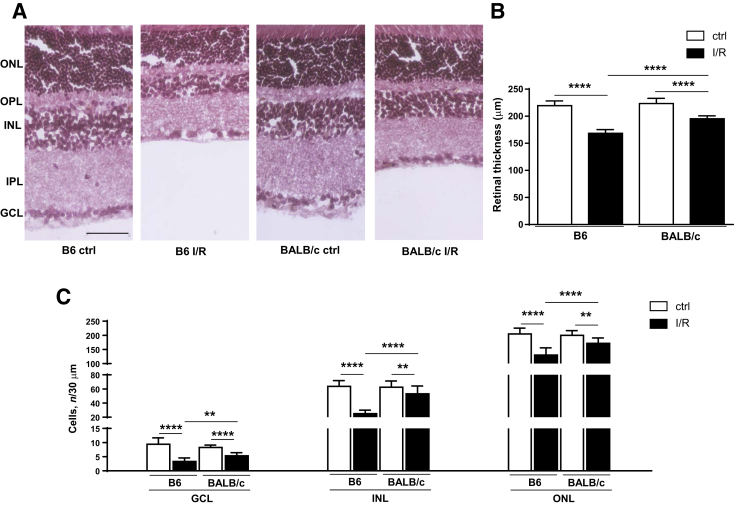
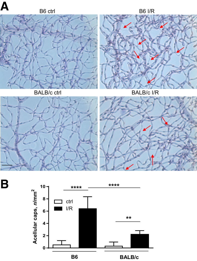
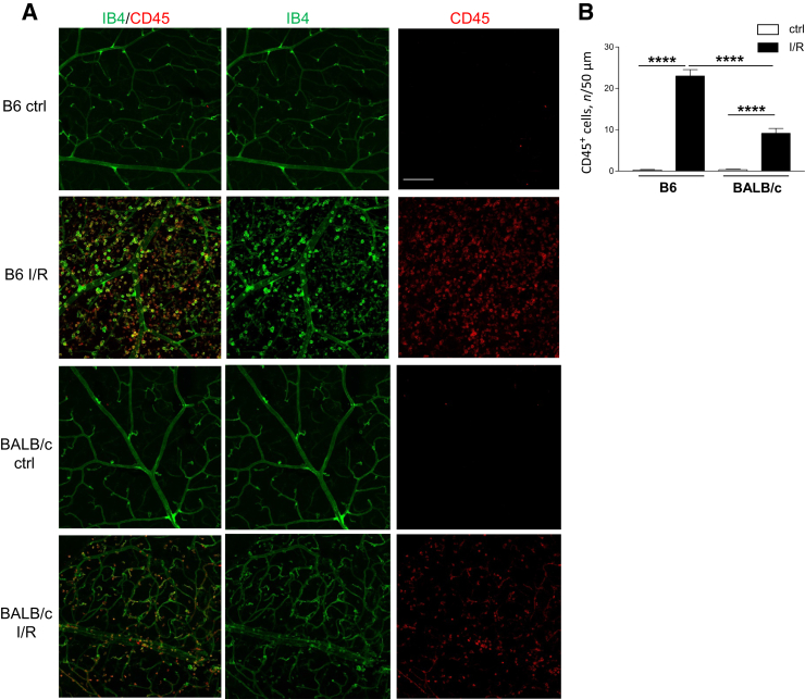
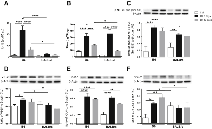
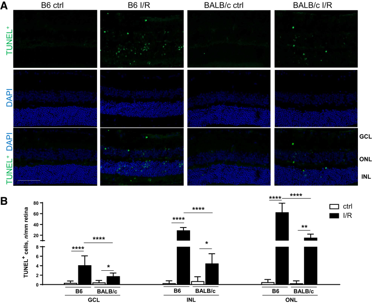
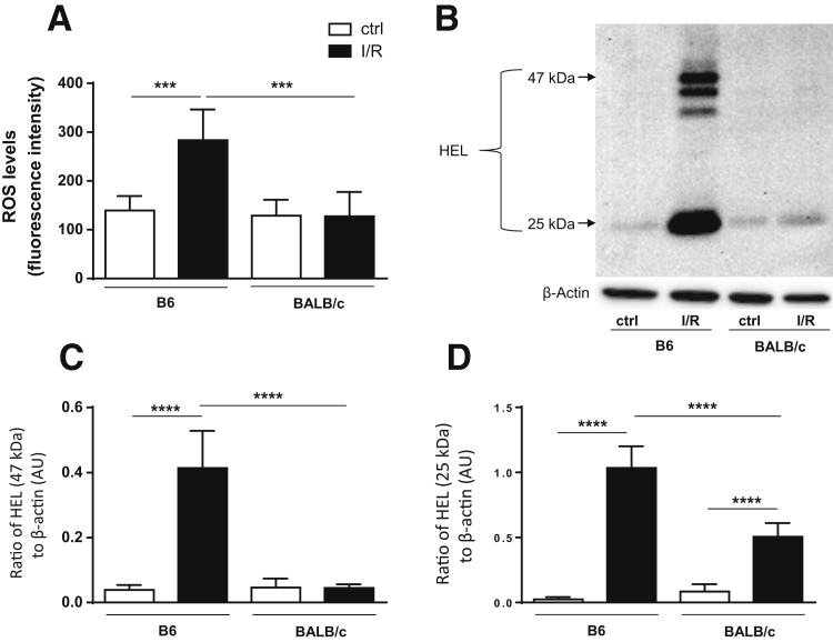
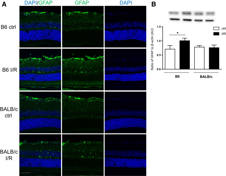
Ischemia is associated with the pathogenesis of retinal disease, including diabetic retinopathy and glaucoma. As a result, the retinal ischemia/reperfusion injury model has been used to study neurovascular changes. Historically, murine models of retinal disease are established in C57BL/6J (B6) mice, which have been described as type 1-dominant responders. In bacterial keratitis models, B6 mice are susceptible, whereas BALB/cJ (BALB/c; type 2-dominant) mice exhibit a resistant phenotype. As such, we questioned whether the type 1/type 2 paradigm could be extrapolated to events associated with retinal pathogenesis. The current study compares the retinal response of B6 with BALB/c mice to investigate strain-specific differences. Retinas were collected at 2 and 10 days after ischemia/reperfusion injury to examine differences in neurovascular degeneration, leukostasis, oxidative stress, glial activation, and select inflammatory mediators. Although both strains showed signs of retinal injury, significantly more damage was observed in B6 mice. Retinal thickness was reduced and vascular damage was more severe in B6 mice. Exacerbated response to injury in B6 versus BALB/c retinas was further supported by increased leukostasis, inflammatory mediators, reactive oxygen species, and lipid peroxidation. In addition, more terminal deoxynucleotidyl transferase-mediated dUTP nick-end labeling-positive cells and increased glial activation were detected in B6 mice. These data indicate that B6 and BALB/c retinas differentially respond to injury, which has broader implications regarding the development and study of retinal diseases.


Figures

Figure 1
Figure 1
B6 retina exhibits significantly increased neurodegeneration compared with BALB/c retina after ischemia/reperfusion (I/R) injury. A: Retinal morphology with hematoxylin/eosin staining isolated from B6 and BALB/c eyes under control (ctrl)/normal conditions and at 2 days after I/R injury. B: Quantitative changes in retinal thickness. C: Quantitative changes in retinal ganglion cell layer (GCL), inner nuclear layer (INL), and outer nuclear layer (ONL). Outer plexiform layer (OPL) and inner plexiform layer (IPL) are also indicated. Data are expressed as means ± SD (B). n = 5 independent experiments (B). ∗∗P < 0.01, ∗∗∗∗P < 0.0001. Scale bar = 50 μm (A, all images).
Figure 2
Figure 2
B6 retinas show significantly more microvascular degeneration compared with BALB/c retinas after ischemia/reperfusion (I/R) injury. A: Isolated retinal microvasculature was stained with hematoxylin-periodic acid-Schiff to detect degenerated capillaries (red arrows) in B6 and BALB/c retinas of normal/control (ctrl) eyes and at 10 days after I/R injury. B: Quantification of acellular capillaries per mm2. Data are expressed as means ± SD (B). n = 6 independent experiments (B). ∗∗P < 0.01, ∗∗∗∗P < 0.0001. Scale bar = 50 μm (A, all images).
Figure 3
Figure 3
Increased leukostasis in B6 ischemia/reperfusion (I/R)–injured retinas compared with BALB/c retinas. A: Retinal flat mounts were isolated from B6 and BALB/c normal controls (ctrls) and at 2 days after injury, then stained with antibodies to CD45 (red) and isolectin B4 (IB4; green). Representative images are provided, showing IB4+ retinal microvasculature and CD45+ leukocytes. B: Quantification of CD45+ cells is presented in the bar graph. n = 5 independent experiments (B). ∗∗∗∗P < 0.0001. Scale bar = 50 μm (A, all images).
Figure 4
Figure 4
Increased inflammation detected in B6 versus BALB/c retinas after ischemia/reperfusion (I/R) injury. Retinal lysates were processed under normal conditions and at 2 and 10 days after injury for protein levels of select proinflammatory mediators: IL-1β (A), tumor necrosis factor (TNF)-α (B), phosphorylated NF-κB p65 at Ser-536 (C), vascular endothelial growth factor (VEGF; D), intracellular adhesion molecule-1 (ICAM-1; E), and cyclooxygenase-2 (COX-2; F). One representative gel image is provided for each Western blot. Data are expressed as means ± SD (A–F). n = 5 independent experiments (A–F). P < 0.05, ∗∗P < 0.01, ∗∗∗P < 0.001, and ∗∗∗∗P < 0.0001. AU, arbitrary unit; Ctrl, control.
Figure 5
Figure 5
Significantly more terminal deoxynucleotidyl transferase-mediated dUTP nick-end labeling–positive (TUNEL+) cells are present in B6 ischemia/reperfusion (I/R)–injured eyes compared with BALB/c eyes. A: Representative images of TUNEL staining in the retina of B6 and BALB/c mice under normal/control (ctrl) conditions and at 2 days after I/R injury. Retinal sections were stained with TUNEL (green) and DAPI (blue). B: Quantification of TUNEL+ cells in ganglion cell layer (GCL), inner nuclear layer (INL), and outer nuclear layer (ONL) is presented in the bar graph. Data are expressed as means ± SD (B). n = 5 independent experiments (B). P < 0.05, ∗∗P < 0.01, and ∗∗∗∗P < 0.0001. Scale bar = 50 μm (A, all images).
Figure 6
Figure 6
Retinas of B6 mice show more reactive oxygen species (ROS) and lipid peroxidation after ischemia/reperfusion (I/R) injury compared with BALB/c mice. Retinal lysates were collected from B6 and BALB/c normal controls (ctrls) and at 2 days after I/R injury. A: ROS levels were determined by fluorescent values. Fluorescent values were acquired by subtraction of negative control values from 2′,7′-dichlorofluorescin diacetate values. B: Lipid peroxidation was determined by Western blot detection of hexanoyl-lysine adduct (HEL) formation in retinal lysates. C: Densitometric analysis of HEL formed at approximately 47 kDa. D: Densitometric analysis of HEL formed at approximately 25 kDa. Data are expressed as means ± SD (A, C, and D). n = 5 independent experiments (A, C, and D). ∗∗∗P < 0.001, ∗∗∗∗P < 0.0001. AU, arbitrary unit.
Figure 7
Figure 7
Retinal stress marker glial fibrillary acidic protein (GFAP) is significantly increased in B6 ischemia/reperfusion (I/R)–injured eye. A: Representative images of GFAP (green) and DAPI (blue) staining in B6 and BALB/c retinal sections under normal conditions and at 2 days after I/R injury. B: Levels of GFAP detected by Western blot analysis in retinal lysates. Data are expressed as means ± SD (B). n = 5 independent experiments (B). P < 0.05. Scale bar = 50 μm (A, all images). AU, arbitrary unit; ctrl, control.

Similar articles

Cited by

References

    1. Bresnick G.H., Engerman R., Davis M.D., de Venecia G., Myers F.L. Patterns of ischemia in diabetic retinopathy. Trans Sect Ophthalmol Am Acad Ophthalmol Otolaryngol. 1976;81:OP694–OP709. - PubMed
    1. Zheng L., Gong B., Hatala D.A., Kern T.S. Retinal ischemia and reperfusion causes capillary degeneration: similarities to diabetes. Invest Ophthalmol Vis Sci. 2007;48:361–367. - PubMed
    1. Hartsock M.J., Cho H., Wu L., Chen W.J., Gong J., Duh E.J. A mouse model of retinal ischemia-reperfusion injury through elevation of intraocular pressure. J Vis Exp. 2016 (113):54065. - PMC - PubMed
    1. Flaherty J.T. Myocardial injury mediated by oxygen free radicals. Am J Med. 1991;91:79S–85S. - PubMed
    1. Kazui M., Andreoni K.A., Williams G.M., Perler B.A., Bulkley G.B., Beattie C., Donham R.T., Sehnert S.S., Burdick J.F., Risby T.H. Visceral lipid peroxidation occurs at reperfusion after supraceliac aortic cross-clamping. J Vasc Surg. 1994;19:473–477. - PubMed

Publication types

Substances