Review
. 2018 Nov;22(11):1011-1025.
doi: 10.1016/j.tics.2018.08.006. Epub 2018 Sep 17.

The Regulatory Role of the Human Mediodorsal Thalamus

Affiliations
Review

The Regulatory Role of the Human Mediodorsal Thalamus

Giulio Pergola et al. Trends Cogn Sci. 2018 Nov.

Abstract

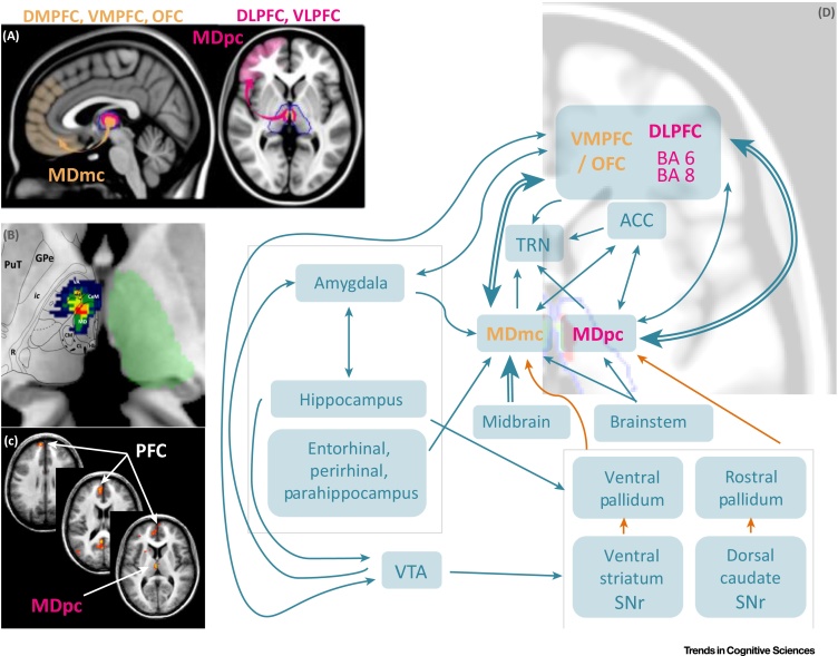
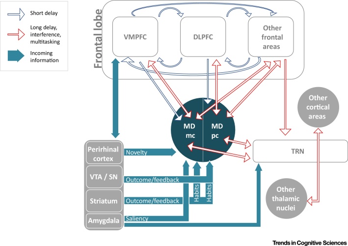
The function of the human mediodorsal thalamic nucleus (MD) has so far eluded a clear definition in terms of specific cognitive processes and tasks. Although it was at first proposed to play a role in long-term memory, a set of recent studies in animals and humans has revealed a more complex, and broader, role in several cognitive functions. The MD seems to play a multifaceted role in higher cognitive functions together with the prefrontal cortex and other cortical and subcortical brain areas. Specifically, we propose that the MD is involved in the regulation of cortical networks especially when the maintenance and temporal extension of persistent activity patterns in the frontal lobe areas are required.

Keywords: mediodorsal thalamus; memory; neuroimaging; persistent activity; prefrontal cortex; temporal extension.


Figures

Figure 1
Figure 1
Connectivity of the MD in Primates. (A) On the left side: connections between the magnocellular MD and the ventromedial and orbitofrontal areas of the PFC. On the right side: connections between the parvocellular MD and the lateral PFC. (B) Overlap of the medial lesions involving the MD of 12 stroke patients (modified from [36]). (C) fMRI activation maps involving the MD and the PFC during episodic retrieval in healthy subjects (modified from [73]). (D) Diagram showing the structural connectivity of the magnocellular MD and the parvocellular MD. Blue arrows correspond to excitatory connections. Orange arrows correspond to inhibitory connections. Abbreviations: ACC, anterior cingulate cortex; CeM, central medial thalamic nucleus; CL, central lateral thalamic nucleus; CM, centromedian thalamic nucleus; DLPFC, dorsolateral prefrontal cortex; DMPFC, dorsomedial prefrontal cortex; GPe, external globus pallidus; Hb, habenula; Ic, internal capsule; MD, mediodorsal thalamic nucleus; MDmc, magnocellular subdivision of the mediodorsal thalamic nucleus; MDpc, parvocellular subdivision of the mediodorsal thalamic nucleus; mtt, mammillothalamic tract; OFC, orbitofrontal cortex; PFC, prefrontal cortex; PuT, putamen; R, thalamic reticular nucleus; SNr, substantia nigra pars reticulata; TRN, thalamic reticular nucleus; VA, ventral–anterior thalamic nucleus; VLPFC, ventrolateral prefrontal cortex; VMPFC, ventromedial prefrontal cortex; VTA, ventral tegmental area.
Figure 2
Figure 2
Key Figure: Regulation of the Prefrontal Cortex Blue contour arrows depict the interactions critical at short delays. In this case, prefrontal reverberation circuits within and across cortical layers may be sufficient for task performance and the interaction with the mediodorsal thalamic nucleus (MD) is represented as an efference copy. Red contour arrows illustrate the hypothesized role of the mediodorsal thalamic nucleus in conditions characterized by long delays, interference, and multitasking. In this case, the prefrontal reverberating circuits depend on back-projections from the MD, which in turn also integrates information coming from several different pathways (turquoise arrows). The MD may be able to recruit further cortical regions by interacting with the thalamic reticular nucleus, which project to all thalamic nuclei, each connected with specific regions of the cortex. Abbreviations: DLPFC, dorsolateral prefrontal cortex; MDmc, magnocellular subdivision of the mediodorsal thalamic nucleus; MDpc, parvocellular subdivision of the mediodorsal thalamic nucleus; SN, substantia nigra; TRN, thalamic reticular nucleus; VMPFC, ventromedial prefrontal cortex; VTA, ventral tegmental area.

Similar articles

Cited by

References

    1. Aggleton J.P., Brown M.W. Episodic memory, amnesia, and the hippocampal-anterior thalamic axis. Behav. Brain Sci. 1999;22:425–444. - PubMed
    1. Golden E.C. Mediodorsal nucleus and its multiple cognitive functions. Neurology. 2016;87:2161–2168. - PubMed
    1. Furtak S.C. Functional neuroanatomy of the parahippocampal region in the rat: the perirhinal and postrhinal cortices. Hippocampus. 2007;17:709–722. - PubMed
    1. Alexander G.E., Fuster J.M. Effects of cooling prefrontal cortex on cell firing in the nucleus medialis dorsalis. Brain Res. 1973;61:93–105. - PubMed
    1. Fuster J.M., Alexander G.E. Neuron activity related to short-term memory. Science. 1971;173:652–654. - PubMed

MeSH terms