. 2018 Nov 16;293(46):17985-17996.
doi: 10.1074/jbc.RA118.004561. Epub 2018 Sep 20.

A partial reconstitution implicates DltD in catalyzing lipoteichoic acid d-alanylation

Affiliations

A partial reconstitution implicates DltD in catalyzing lipoteichoic acid d-alanylation

B McKay Wood et al. J Biol Chem. .

Abstract

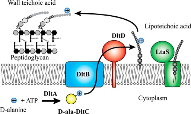
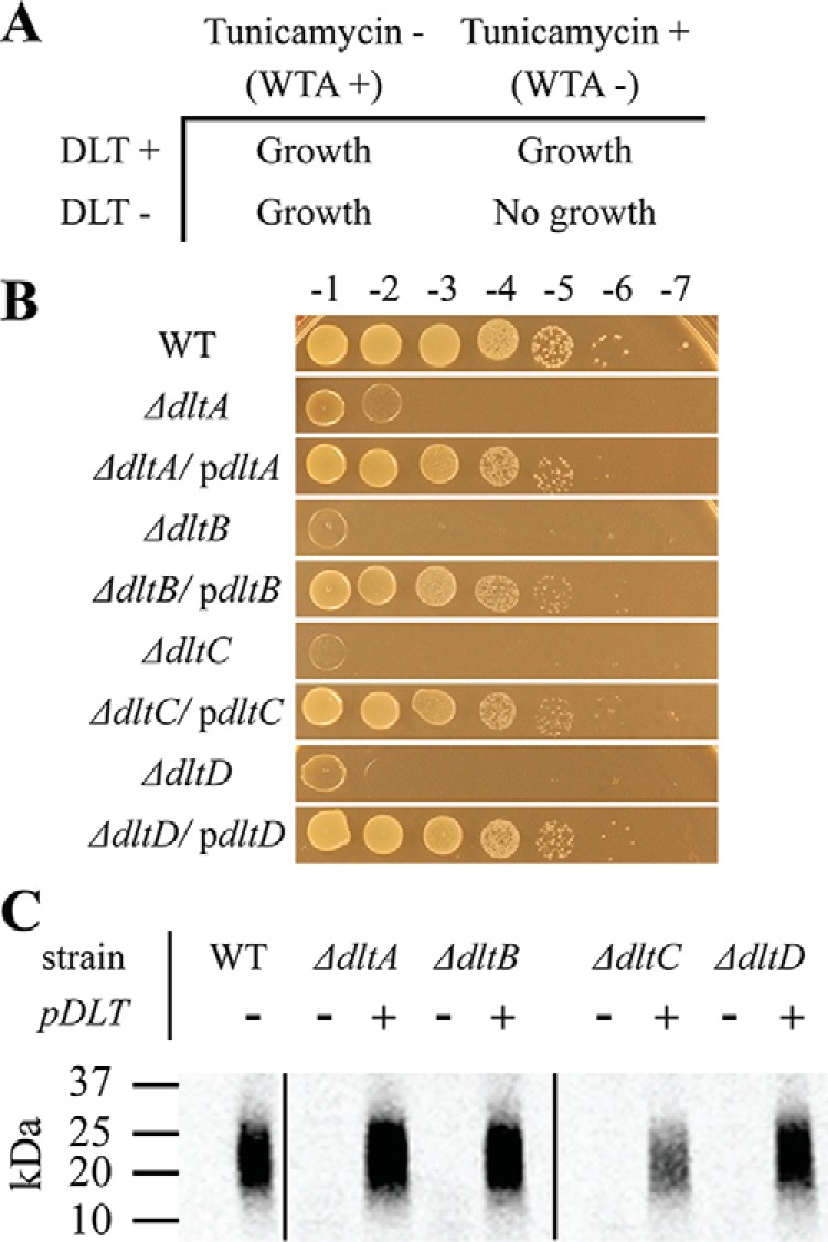
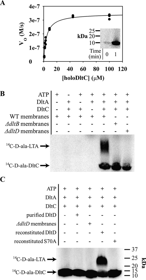
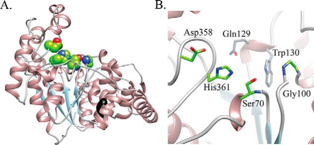
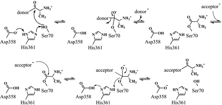
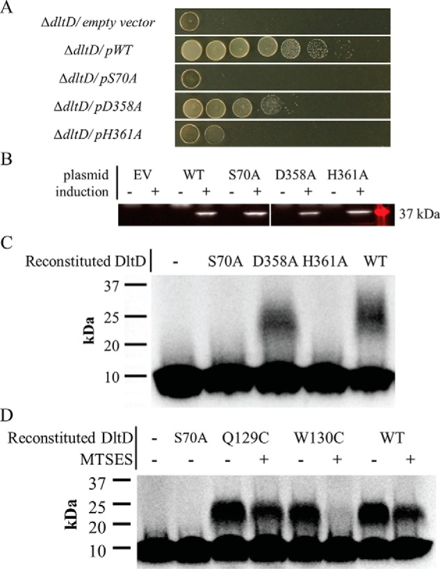
Modifications to the Gram-positive bacterial cell wall play important roles in antibiotic resistance and pathogenesis, but the pathway for the d-alanylation of teichoic acids (DLT pathway), a ubiquitous modification, is poorly understood. The d-alanylation machinery includes two membrane proteins of unclear function, DltB and DltD, which are somehow involved in transfer of d-alanine from a carrier protein inside the cell to teichoic acids on the cell surface. Here, we probed the role of DltD in the human pathogen Staphylococcus aureus using both cell-based and biochemical assays. We first exploited a known synthetic lethal interaction to establish the essentiality of each gene in the DLT pathway for d-alanylation of lipoteichoic acid (LTA) and confirmed this by directly detecting radiolabeled d-Ala-LTA both in cells and in vesicles prepared from mutant strains of S. aureus We developed a partial reconstitution of the pathway by using cell-derived vesicles containing DltB, but no other components of the d-alanylation pathway, and showed that d-alanylation of previously formed lipoteichoic acid in the DltB vesicles requires the presence of purified and reconstituted DltA, DltC, and DltD, but not of the LTA synthase LtaS. Finally, based on the activity of DltD mutants in cells and in our reconstituted system, we determined that Ser-70 and His-361 are essential for d-alanylation activity, and we propose that DltD uses a catalytic dyad to transfer d-alanine to LTA. In summary, we have developed a suite of assays for investigating the bacterial DLT pathway and uncovered a role for DltD in LTA d-alanylation.

Keywords: DLT pathway; DltB; MBOAT; Staphylococcus aureus (S. aureus); acyltransferase; cell wall; membrane protein; pathway reconstitution; teichoic acid.


Conflict of interest statement

The authors declare that they have no conflicts of interest with the contents of this article

Figures

Figure 1.
Figure 1.
Model for teichoic acid d-alanylation by the DLT pathway. DltA-catalyzed DltC d-alanylation takes place in the cytoplasm at the expense of ATP. What happens to the thioesterified d-Ala-DltC is unclear, but the two predicted membrane-bound acyltransferases, DltB and DltD, are involved in the migration of the d-alanyl moiety across the membrane and onto lipoteichoic acid and wall teichoic acid. Lipoteichoic acid is a poly(glycerol phosphate) polymer that is biosynthesized on a diglucosyl diacylglycerol lipid anchor outside the cell by LtaS.
Figure 2.
Figure 2.
DltABCD are each necessary for LTA d-alanylation. The synthetic lethal interaction between the DLT pathway and WTA biosynthesis pathway allows for dlt selection. Our laboratory previously reported that the natural product, tunicamycin, is a potent inhibitor of TarO in WTA biosynthesis. A illustrates how either TarO inhibition or genetic DLT inactivation alone do not hinder growth, whereas tunicamycin treatment of a dlt-null mutant inhibits bacterial growth. WT S. aureus and null mutants of individual dlt genes were transformed with either plasmid-encoded dltABCD or empty vector. In B, growth on 0.4 μg/ml tunicamycin was used as a readout of DLT activity. The growth dependence of each null mutant on a plasmid-borne copy of the dlt operon demonstrates the essentiality of their respective dlt gene for teichoic acid d-alanylation. To directly detect changes in LTA d-alanylation, the same strains were fed d-[14C]alanine before SDS extraction, SDS-PAGE, and autoradiography as described under “In vivo LTA d-alanylation assay.” C shows the level of d-Ala-LTA produced in each mutant with or without complementation. d-Ala-LTA migrates as a smear between 20 and 25 kDa as compared with protein ladder.
Figure 3.
Figure 3.
In vitro DLT activity assays show essentiality of each DLT protein for d-alanylation. A continuous ATP-hydrolysis assay for DltA-catalyzed DltC d-alanylation was developed using NADH turnover via a three-enzyme system for ATP regeneration (adenylate kinase, pyruvate kinase, and lactate dehydrogenase). A shows nonlinear curve fitting of V0 with varying concentrations of holoDltC plotted with the Michaelis–Menten equation. Three data sets are shown plotted together. DltA assay conditions were modified slightly for in vitro d-[14C]alanine incorporation into DltC (inset to A). Next, vesicles composed of fractionated membranes from S. aureus and dlt-null mutant cells were incubated with d-[14C]Ala-DltC. The left four lanes in B show that d-[14C]Ala-DltC was produced only when ATP, DltA, and DltC were all added, and no native DltC was contributed from the vesicle preparation. Using vesicles prepared with membranes from dltB- and dltD-null strains, the right three lanes of B show that d-[14C]Ala-LTA formation depends on both DltB and DltD. Lastly, the full-length DltD enzyme was purified and reconstituted into vesicles. The gel in C shows that d-[14C]Ala-LTA formation in vesicles from dltD-null membranes required DltD reconstitution and that an inactivated point mutant of DltD failed to support the same activity when reconstituted.
Figure 4.
Figure 4.
Homology model of S. aureus DltD demonstrates a Ser-His-Asp triad. The Phyre 2–calculated model of the S. aureus DltD is shown in A. An unpublished crystal structure of the ortholog from S. pneumoniae without the transmembrane helix was used as a template (PDB code 3BMA). The enzyme forms an αβα sandwich-like fold, and the black colored ribbon indicates where the N-terminal transmembrane helix would originate (before Glu-33). Structural similarity searches of the Protein Data Bank retrieved several families of SGNH hydrolase-like proteins (Fig. S11). The Ser-His-Asp triad (green sphere representation in A) in the DltD active site has been identified as a catalytic triad in SGNH hydrolases. In the active-site zoomed image in B, the amide backbones of Ser-70 and Gly-100 are well-positioned for oxyanion stabilization, and the conserved DltD residues Gln-129 and Trp-130 shown in gray appear to form the back of the pocket.
Scheme 1.
Scheme 1.
Proposed mechanism for acyl transfer via DltD's catalytic dyad.
Figure 5.
Figure 5.
DltD-dependent LTA d-alanylation depends on an active-site pocket that includes Ser-70, Trp-130, His-361, and Asp-358. Site-directed alanine substitutions of DltD's apparent catalytic triad in an S. aureus expression vector were transformed into the S. aureus dltD-null strain. A shows a test of their activity via growth complementation in the presence of 0.4 μg/ml tunicamycin. Serial dilutions were spotted on agar plates from undiluted (left) to 10−7 diluted (right). Here, “−” indicates empty vector (EV), and WT, S70A, D358A, and H361A refer to the myc-dltD alleles expressed as part of plasmid-encoded dltABCD. Western blotting to the N-terminal myc tags fused to each DltD clone allowed confirmation of their expression (B). The tag showed no effect on DltD function (Fig. S15). Then, the mutant DltD enzymes were expressed in E. coli, purified, and reconstituted into vesicles made from S. aureus dltD-null membranes. C shows the result of in vitro LTA d-alanylation assay of these reconstituted DltD vesicles using d-[14C]Ala-DltC. Here, “−” refers to vesicles without DltD reincorporation. Lastly, in panel D, the Q129C and W130C were each tested for their ability to support LTA d-alanylation in vitro with or without the presence of thiol-reactive MTSES. Purified DltD WT and mutants were reconstituted into vesicles of membranes from the S. aureus dltD-null mutant. The reconstituted vesicles were pretreated with 5 mm MTSES where indicated with a “+.” The inactive S70A mutant-reconstituted vesicles as well as vesicles with no DltD added were run in the first two lanes for comparison.

Similar articles

Cited by

References

    1. Weidenmaier C., and Peschel A. (2008) Teichoic acids and related cell-wall glycopolymers in Gram-positive physiology and host interactions. Nat. Rev. Microbiol. 6, 276–287 10.1038/nrmicro1861 - DOI - PubMed
    1. Percy M. G., and Gründling A. (2014) Lipoteichoic acid synthesis and function in Gram-positive bacteria. Annu. Rev. Microbiol. 68, 81–100 10.1146/annurev-micro-091213-112949 - DOI - PubMed
    1. Brown S., Santa Maria J. P. Jr., and Walker S. (2013) Wall teichoic acids of Gram-positive bacteria. Annu. Rev. Microbiol. 67, 313–336 10.1146/annurev-micro-092412-155620 - DOI - PMC - PubMed
    1. Neuhaus F. C., and Baddiley J. (2003) A continuum of anionic charge: structures and functions of d-alanyl-teichoic acids in Gram-positive bacteria. Microbiol. Mol. Biol. Rev. 67, 686–723 10.1128/MMBR.67.4.686-723.2003 - DOI - PMC - PubMed
    1. Kovács M., Halfmann A., Fedtke I., Heintz M., Peschel A., Vollmer W., Hakenbeck R., and Brückner R. (2006) A functional dlt operon, encoding proteins required for incorporation of d-alanine in teichoic acids in Gram-positive bacteria, confers resistance to cationic antimicrobial peptides in Streptococcus pneumoniae. J. Bacteriol. 188, 5797–5805 10.1128/JB.00336-06 - DOI - PMC - PubMed

Publication types

MeSH terms

Associated data

LinkOut - more resources