. 2018 Nov 23;293(47):18055-18070.
doi: 10.1074/jbc.RA118.005439. Epub 2018 Sep 20.

The properties of Msh2-Msh6 ATP binding mutants suggest a signal amplification mechanism in DNA mismatch repair

Affiliations

The properties of Msh2-Msh6 ATP binding mutants suggest a signal amplification mechanism in DNA mismatch repair

William J Graham 5th et al. J Biol Chem. .

Abstract

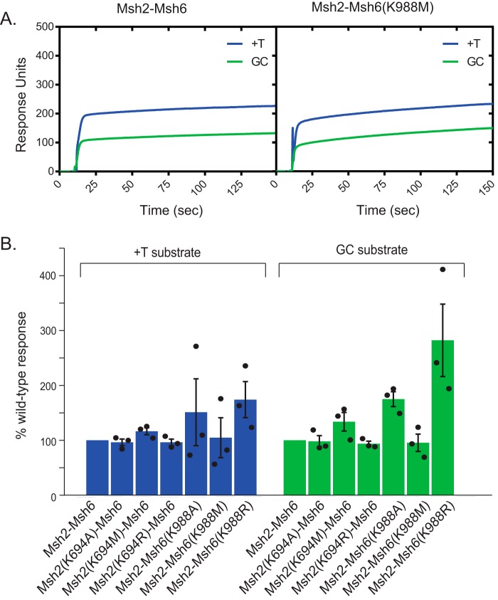
DNA mismatch repair (MMR) corrects mispaired DNA bases and small insertion/deletion loops generated by DNA replication errors. After binding a mispair, the eukaryotic mispair recognition complex Msh2-Msh6 binds ATP in both of its nucleotide-binding sites, which induces a conformational change resulting in the formation of an Msh2-Msh6 sliding clamp that releases from the mispair and slides freely along the DNA. However, the roles that Msh2-Msh6 sliding clamps play in MMR remain poorly understood. Here, using Saccharomyces cerevisiae, we created Msh2 and Msh6 Walker A nucleotide-binding site mutants that have defects in ATP binding in one or both nucleotide-binding sites of the Msh2-Msh6 heterodimer. We found that these mutations cause a complete MMR defect in vivo The mutant Msh2-Msh6 complexes exhibited normal mispair recognition and were proficient at recruiting the MMR endonuclease Mlh1-Pms1 to mispaired DNA. At physiological (2.5 mm) ATP concentration, the mutant complexes displayed modest partial defects in supporting MMR in reconstituted Mlh1-Pms1-independent and Mlh1-Pms1-dependent MMR reactions in vitro and in activation of the Mlh1-Pms1 endonuclease and showed a more severe defect at low (0.1 mm) ATP concentration. In contrast, five of the mutants were completely defective and one was mostly defective for sliding clamp formation at high and low ATP concentrations. These findings suggest that mispair-dependent sliding clamp formation triggers binding of additional Msh2-Msh6 complexes and that further recruitment of additional downstream MMR proteins is required for signal amplification of mispair binding during MMR.

Keywords: DNA binding protein; DNA endonuclease; DNA mismatch repair; DNA repair; DNA replication; Mlh1-Pms1; Msh2-Msh6; MutS; S. cerevisiae; exonuclease 1; mutagenesis.


Conflict of interest statement

The authors declare that they have no conflicts of interest with the contents of this article

Figures

Figure 1.
Figure 1.
Effects of mutations on the ability of mutant subunits to bind ATP. A, ATP-binding site from E. coli MutS (Protein Data Bank (PDB) code 5akb (29)) depicting highly conserved residues from the Walker A motif (Lys694 in S. cerevisiae Msh2 and Lys988 in S. cerevisiae Msh6) and their position relative to an AMP-PNP ATP analog. B, histogram showing relative Msh2 binding of 60 μm [γ-32P]ATP under nonhydrolyzable conditions. C, histogram showing relative Msh6 binding of 1 μm [γ-32P]ATP under nonhydrolyzable conditions. Error bars represent the S.D. from the mean, and individual values from different experiments are indicated by the dots on each histogram bar.
Figure 2.
Figure 2.
Msh2 and Msh6 mutants are partially defective in an in vitro reconstituted MMR assay. A, schematic representation of the DNA substrate for the reconstituted repair assay. Substrates had a nick at either the NaeI or AflIII site but not both. B, representative gel showing repair of the 5′ nicked NaeI substrate used in an assay with 2.5 mm ATP. C–F, the amount of repair relative to WT Msh2–Msh6 was quantitated for the 5′ nicked NaeI substrate at 2.5 mm ATP (100% repair = 43.5% of substrate repaired) (C), the 5′ nicked NaeI substrate at 0.1 mm ATP (100% repair = 17.8% of substrate repaired) (D), the 3′ nicked AflIII substrate at 2.5 mm ATP (100% repair = 23.2% of substrate repaired) (E), or the 3′ nicked AflIII substrate at 0.1 mm ATP (100% repair = 12.1% of substrate repaired) (F) by normalizing the percentage of repair in each experiment to the WT levels and averaging a minimum of at least three independent experiments. Error bars represent the S.D. from the mean, and individual values from different experiments are indicated by the dots on each histogram bar. In F, numbers over the histogram bars are the average of the ratio of mutant repair to repair without Msh2–Msh6 (compared internally between experiments) with the error indicating the S.D.
Figure 3.
Figure 3.
Analysis of Msh2–Msh6-mispair binding in the absence of nucleotide. A, representative SPR sensorgrams showing WT Msh2–Msh6 or Msh2–Msh6(K988M) binding to mispaired (+T; blue) or base-paired (GC; green) DNA in the absence of ATP. B, plot of the percentage of WT Msh2–Msh6 steady-state response (RUmax) for the +T mispair–containing substrate (left, blue) and the GC fully base-paired control substrate (right, green); the level of binding was normalized to the WT level of binding, which was set at 100%. In all cases, the R2 values for the fits to determine RUmax were above 0.993, and in most cases, the R2 values for the fits were above 0.999. Error bars represent the S.D. from the mean, and individual values from different experiments are indicated by the dots on each histogram bar.
Figure 4.
Figure 4.
Msh2 and Msh6 mutants have mispair binding defects in the presence of ATP and are affected by ATP differentially from WT Msh2–Msh6. A, representative sensorgram showing substantial or little sliding clamp formation by WT Msh2–Msh6, Msh2(K694M)–Msh6, and Msh2–Msh6(K988M) on +T mispair–containing substrate (blue) but little sliding clamp formation on fully base-paired substrate (green). Msh2–Msh6 was added at 10 s, and IPTG was added at 160 s. B, fitted steady-state response (RUmax) of WT Msh2–Msh6 (red) and Msh2–Msh6(K988M) (purple) on a +T mispair–containing substrate is plotted as a function of ATP concentration. In all cases, fits to the association curves had R2 values greater than 0.996. C, the molar ratio of Msh2–Msh6 complexes to DNA at steady state (RUmax) was calculated for WT and mutant Msh2–Msh6 complexes at different ATP concentrations. In all cases, fits to the association curves had R2 values greater than 0.996. D, the percentage of Msh2–Msh6 dissociation for WT Msh2–Msh6 (red) and Msh2–Msh6(K988M) (purple) at 200 s (corresponding to 40 s or >10 half-lives for the WT sliding clamp after the addition of IPTG) is plotted as a function of ATP concentration. E, the percentage of Msh2–Msh6 dissociation was determined for WT and mutant Msh2–Msh6 complexes at different ATP concentrations. In C and E, individual values from different experiments are indicated by the dots on each histogram bar.
Figure 5.
Figure 5.
Analysis of Msh2–Msh6 directly dissociating from double end–blocked DNA. A, representative surface plasmon resonance sensorgrams showing WT and mutant Msh2–Msh6 binding to, and directly dissociating from, mispaired DNA in the presence of ATP. Direct dissociation reactions (red and purple curves) were performed the same as the sliding clamp formation experiments except IPTG and Msh2–Msh6 were omitted from the ATP wash step. Consequently, any Msh2–Msh6 leaving the DNA must do so directly due to the continual presence of the LacI end block. B, half-lives of the major component of dissociation of WT and mutant Msh2–Msh6 complexes from double end–blocked DNA. In all cases, fits to the dissociation curves had R2 values greater than 0.997.
Figure 6.
Figure 6.
Msh2 and Msh6 mutants can recruit Mlh1–Pms1 to mispaired DNA. A, representative SPR sensorgram for Mlh1–Pms1 recruitment on GT mispair–containing DNA by WT Msh2–Msh6 (red) or Msh2–Msh6(K988M) (purple); Mlh1–Pms1 addition is indicated by the rightmost vertical dotted line. B, molar ratios were calculated by determining the increase in RUmax due to Msh2–Msh6 and Mlh1–Pms1 from fits to the association curves (see “Experimental procedures”) and by scaling these responses by the molecular weights of the Msh2–Msh6 and the Mlh1–Pms1 complexes. In all cases, fits to the association curves had R2 values greater than 0.998. Error bars represent the S.D. from the mean, and individual values from different experiments are indicated by the dots on each histogram bar.
Figure 7.
Figure 7.
Msh2 and Msh6 mutants are partially defective for Mlh1–Pms1 endonuclease activation. Assays contain WT or mutant Msh2–Msh6, Mlh1–Pms1, PCNA, RFC-Δ1N, a circular DNA substrate with a mispair and nick 3′ to the mispair at the AflIII site (see Fig. 2A for schematic of DNA substrate), and 0.1 or 2.5 mm ATP. A, representative Southern blot showing levels of nicking promoted by WT or mutant Msh2–Msh6 at 2.5 mm ATP. B and C, quantitation of reactions containing 2.5 mm ATP (100% nicking = 22.6% of substrate nicked) (B) and 0.1 mm ATP (100% nicking = 17.2% of substrate nicked) (C) were performed by normalizing the percentage of repair in each experiment to the WT levels and averaging a minimum of at least three independent experiments. Error bars represent the S.D. from the mean, and individual values from different experiments are indicated by the dots on each histogram bar. In C, numbers over the histogram bars are the average of the ratio of mutant nicking to nicking without Msh2–Msh6 (compared internally between experiments) with the error indicating the S.D.

Similar articles

Cited by

References

    1. Fishel R. (2015) Mismatch repair. J. Biol. Chem. 290, 26395–26403 10.1074/jbc.R115.660142 - DOI - PMC - PubMed
    1. Li Z., Pearlman A. H., and Hsieh P. (2016) DNA mismatch repair and the DNA damage response. DNA Repair 38, 94–101 10.1016/j.dnarep.2015.11.019 - DOI - PMC - PubMed
    1. Kolodner R. D., and Marsischky G. T. (1999) Eukaryotic DNA mismatch repair. Curr. Opin. Genet. Dev. 9, 89–96 10.1016/S0959-437X(99)80013-6 - DOI - PubMed
    1. Li G. M. (2008) Mechanisms and functions of DNA mismatch repair. Cell Res. 18, 85–98 10.1038/cr.2007.115 - DOI - PubMed
    1. Spies M., and Fishel R. (2015) Mismatch repair during homologous and homologous recombination. Cold Spring Harb. Perspect. Biol. 7, a022657 10.1101/cshperspect.a022657 - DOI - PMC - PubMed

Publication types

MeSH terms

Associated data