. 2018 Nov 2;293(44):17267-17277.
doi: 10.1074/jbc.RA118.004461. Epub 2018 Sep 20.

Quaternary structure of the small amino acid transporter OprG from Pseudomonas aeruginosa

Affiliations

Quaternary structure of the small amino acid transporter OprG from Pseudomonas aeruginosa

Raghavendar Reddy Sanganna Gari et al. J Biol Chem. .

Abstract

Pseudomonas aeruginosa is an opportunistic human pathogen that causes nosocomial infections. The P. aeruginosa outer membrane contains specific porins that enable substrate uptake, with the outer membrane protein OprG facilitating transport of small, uncharged amino acids. However, the pore size of an eight-stranded β-barrel monomer of OprG is too narrow to accommodate even the smallest transported amino acid, glycine, raising the question of how OprG facilitates amino acid uptake. Pro-92 of OprG is critically important for amino acid transport, with a P92A substitution inhibiting transport and the NMR structure of this variant revealing that this substitution produces structural changes in the barrel rim and restricts loop motions. OprG may assemble into oligomers in the outer membrane (OM) whose subunit interfaces could form a transport channel. Here, we explored the contributions of the oligomeric state and the extracellular loops to OprG's function. Using chemical cross-linking to determine the oligomeric structures of both WT and P92A OprG in native outer membranes and atomic force microscopy, and single-molecule fluorescence of the purified proteins reconstituted into lipid bilayers, we found that both protein variants form oligomers, supporting the notion that subunit interfaces in the oligomer could provide a pathway for amino acid transport. Furthermore, performing transport assays with loop-deleted OprG variants, we found that these variants also can transport small amino acids, indicating that the loops are not solely responsible for substrate transport. We propose that OprG functions as an oligomer and that conformational changes in the barrel-loop region might be crucial for its activity.

Keywords: Pseudomonas aeruginosa (P. aeruginosa); atomic force microscopy (AFM); chemical modification; fluorescence; membrane protein; outer membrane; single-molecule biophysics; transporter.


Conflict of interest statement

The authors declare that they have no conflicts of interest with the contents of this article

Figures

Figure 1.
Figure 1.
Structure models of monomeric and hypothetical trimeric OprG. A, top view of the NMR structure of OprG (PDB code 2N6L). Residues pointing inside the barrel are shown as ball and stick models and the solvent accessible surface of the barrel as a gray translucent volume. This clearly indicates that there is no luminal channel large enough to transport small amino acids. B, homology model of a hypothetical OprG trimer based on the crystal structure of OmpW (PDB code 2F1V). The solvent accessible surface shows a channel along the trimeric interface large enough to accommodate the amino acid glycine. C, disulfide-linked trimer of T65C–L90C OprG based on the homology model of B. D, disulfide-linked trimer of S128C–S136C OprG based on the homology model of B.
Figure 2.
Figure 2.
Cross-linking of OprG oligomers in native membranes. A, Western blotting against StrepTag II of outer membrane vesicles from P. aeruginosa PAO1 expressing five different double cysteine mutants of OprG as isolated (2nd lanes) and cross-linked with CuP (1st lanes), BM(PEG)3 (3rd lanes), or oPDM (4th lanes), respectively. Outer membrane vesicles were treated with cross-linker, quenched, boiled in nonreducing, denaturing sample buffer before SDS-PAGE and Western blot analysis. The expected position of unfolded OprG monomers to tetramers are indicated by arrows. B, Coomassie Brilliant Blue stained SDS-PAGE of purified, cross-linked OprG double cysteine mutants. Outer membrane vesicles from P. aeruginosa PAO1 expressing the 128Cys–Cys136 mutant of WT or P92A OprG were cross-linked with oPDM or CuP, respectively, and solubilized in SB 3–14. Cross-linked OprG oligomers were then purified over StrepTactin XT resin and separated by size exclusion chromatography over Superdex 200. Samples were run boiled (+) and non-boiled (−) to assess folding states of the β-barrel. The expected positions of unfolded OprG monomers to tetramers are indicated by arrows. For the reversible cross-linker CuP, samples were also run after reduction to monomers by β-mercaptoethanol (β-ME). Coomassie-stained SDS-PAGE gels are representatives of experiments that were replicated three (independent purifications) to 10 (gels) times. The labels for molecular weight markers (MW) apply to all markers in the rows of A and B.
Figure 3.
Figure 3.
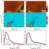
AFM of OprG in lipid bilayers. A, representative image of WT OprG in lipid bilayer. B, representative image of P92A OprG in lipid bilayer. Lipid bilayers were identified by measuring step heights at the edges, which can be seen in both images. Protrusions of the individual OprG particles above the membrane were visualized by AFM (light brown color). The full color scale is shown on the right. C and D, height-volume distributions of membrane protrusions of WT (C) and P92A (D) OprG in lipid bilayers. E and F, modeling of the experimental volume distributions (red) of WT (E) and P92A (F) OprG as superpositions of Gaussians (dashed blue lines). The sums of these Gaussians are shown in black.
Figure 4.
Figure 4.
Single molecule photobleaching of OprG in lipid bilayers. A and B, bleaching steps of single fluorescent protein complexes in lipid bilayers reveal the numbers of Alexa 647-labeled subunits. Examples of single-step (A) and two-step (B) bleaching traces are shown. The black lines represent mean intensity values from 5 × 5 pixel2 regions. The red lines represent the Chung-Kennedy nonlinear filtered versions of the data. C, average fractions of observed bleaching steps from 10 movies of two different protein reconstitutions. Division of bars into blue, green, and red illustrate the calculated fractions of monomers, dimers, and trimers within one observed population, as explained under “Experimental procedures.” D, total oligomeric fractions of monomers (blue), dimers (green), and trimers (red) are calculated from data in C.
Figure 5.
Figure 5.
Amino acid transport of loop deletion mutants of OprG. Liposome-swelling assay with reconstituted loop deletion mutants ΔL1, ΔL2, ΔL3, and ΔL4 of OprG in the presence of amino acids known to be transported by the WT OprG. The data are presented as mean ± S.D. (dashed line) of two to three biological replicates, i.e. repeated purifications and refoldings of loop deletion mutants, each with triplicate permeation measurements. Data for WT OprG (wt) and protein-free liposomes (blank) are shown as positive and negative controls, respectively.

Similar articles

Cited by

References

    1. Strateva T., and Yordanov D. (2009) Pseudomonas aeruginosa: a phenomenon of bacterial resistance. J. Med. Microbiol. 58, 1133–1148 10.1099/jmm.0.009142-0 - DOI - PubMed
    1. Kang C. I., Kim S. H., Kim H. B., Park S. W., Choe Y. J., Oh M. D., Kim E. C., and Choe K. W. (2003) Pseudomonas aeruginosa bacteremia: risk factors for mortality and influence of delayed receipt of effective antimicrobial therapy on clinical outcome. Clin. Infect. Dis. 37, 745–751 10.1086/377200 - DOI - PubMed
    1. Hancock R. E., and Brinkman F. S. (2002) Function of pseudomonas porins in uptake and efflux. Annu. Rev. Microbiol. 56, 17–38 10.1146/annurev.micro.56.012302.160310 - DOI - PubMed
    1. Davies D. (2003) Understanding biofilm resistance to antibacterial agents. Nat. Rev. Drug Discov. 2, 114–122 10.1038/nrd1008 - DOI - PubMed
    1. Hall-Stoodley L., Costerton J. W., and Stoodley P. (2004) Bacterial biofilms: from the natural environment to infectious diseases. Nat. Rev. Microbiol. 2, 95–108 10.1038/nrmicro821 - DOI - PubMed

Publication types

MeSH terms

Substances

LinkOut - more resources