. 2018 Nov 9;132(4):jcs219501.
doi: 10.1242/jcs.219501.

PLK4 is a microtubule-associated protein that self-assembles promoting de novo MTOC formation

Affiliations

PLK4 is a microtubule-associated protein that self-assembles promoting de novo MTOC formation

Susana Montenegro Gouveia et al. J Cell Sci. .

Abstract

The centrosome is an important microtubule-organising centre (MTOC) in animal cells. It consists of two barrel-shaped structures, the centrioles, surrounded by the pericentriolar material (PCM), which nucleates microtubules. Centrosomes can form close to an existing structure (canonical duplication) or de novo How centrosomes form de novo is not known. The master driver of centrosome biogenesis, PLK4, is critical for the recruitment of several centriole components. Here, we investigate the beginning of centrosome biogenesis, taking advantage of Xenopus egg extracts, where PLK4 can induce de novo MTOC formation ( Eckerdt et al., 2011; Zitouni et al., 2016). Surprisingly, we observe that in vitro, PLK4 can self-assemble into condensates that recruit α- and β-tubulins. In Xenopus extracts, PLK4 assemblies additionally recruit STIL, a substrate of PLK4, and the microtubule nucleator γ-tubulin, forming acentriolar MTOCs de novo The assembly of these robust microtubule asters is independent of dynein, similar to what is found for centrosomes. We suggest a new mechanism of action for PLK4, where it forms a self-organising catalytic scaffold that recruits centriole components, PCM factors and α- and β-tubulins, leading to MTOC formation.This article has an associated First Person interview with the first author of the paper.

Keywords: Centrosome; De novo assembly; In vitro reconstitution; MTOCs; Microtubule nucleation; PCM; PLK4; Supramolecular assembly.


Conflict of interest statement

Competing interestsThe authors declare no competing or financial interests.

Figures
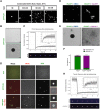
Fig. 1.
Fig. 1.
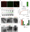
PLK4 self-assembly is dependent on its kinase activity and PLK4 assemblies concentrate tubulin in vitro. (A) Representative confocal images of GFP–PLK4 assemblies formed at different concentrations of NaCl. Scale bars: 2 μm. (B) Electron microscopy (EM) images showing PLK4 assemblies in vitro. Scale bar: 2 μm (top); 100 nm (bottom). (C) Fluorescence intensity recovery after photobleaching (FRAP) (mean±s.d.; %) analysis of PLK4 assemblies in vitro. The inset to the graph shows a magnification of the recovery plot; grey shading indicates ±s.d. Scale bar: 2 μm. (D) Confocal images representing GFP–PLK4AS in the absence or presence of 1-Naphthyl-PP1 (1NA-PP1, PP1 analogue). DMSO was used as a control for 1NA-PP1. Scale bar: 5 µm. Note that, in presence of 1NA-PP1, GFP–PLK4 forms disorganised structures. (E) EM images of GFP–PLK4AS in the presence or absence of 1NA-PP1. Scale bars: 100 nm. (F) Quantification of sphere-like assemblies versus aggregates obtained from EM data. Three independent experiments were counted. (G) Confocal images of GFP–PLK4 assembly formation in the absence or presence of Rhodamine-labelled tubulin (500 nM). GFP was used as a control. Scale bars: 5 µm; insets, 2 µm. (H) FRAP analysis as in C of tubulin coating PLK4 spheres in vitro, showing that they have little dynamicity. Scale bars: 2 μm.
Fig. 2.
Fig. 2.
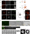
PLK4 is a microtubule-associated protein that promotes microtubule bundling in vitro. (A) Confocal images of Taxol-stabilised MTs alone (Rhodamine-labelled tubulin, red), recombinant purified GFP–PLK4 alone (green) and the mixture of both, showing association of PLK4 condensates to MTs. Scale bar: 5 μm; inset, 2 µm. (B) Quantification of PLK4 assemblies (mean±s.d.; %) associated to MTs compared to free PLK4 in the background (N=3, n=100 spot/condition). (C) MT-pelleting assays. The two assays are showing either a constant PLK4 concentration (0.7 μM) mixed and incubated with different MT concentrations (0 to 4 μM) or increasing amounts of GFP–PLK4 (0 to 4 µM) in the presence of a constant MT concentration (10 µM). The Coomassie Blue-stained gel is showing supernatant (S) and pellet (P) for each condition. (D) Quantitative analysis of binding properties between PLK4 and MTs. Note that the dissociation constant (Kd) for PLK4, determined by best fit to the data (red curve), is 0.62±0.071 μM. Note that the dotted line is the real data (mean±s.e.m.) and the red line is the fitted curve to derive constants. The data were collected from three independent experiments. (E) EM images showing MTs alone or MTs incubated with two concentrations of PLK4 (0.1 µM and 1 µM). Scale bars: 100 nm. (F) Percentage of single or bundled MTs quantified from EM data in presence of PLK4 (0.1 µM or 1 µM); MTs alone are used as a control. Results are mean±s.e.m. scored using 30 images per condition obtained from three independent experiments each (***P<0.001; **P<0.01, Student's t-test). (G) Time course of PLK4 (1 µM) incubated with MTs. Note that PLK4 binds to MTs before PLK4 condensates are formed (“≡” means ∼0 min, as feasible experimentally). Scale bars: 100 nm.
Fig. 3.
Fig. 3.
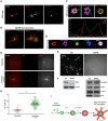
PLK4 condensates form de novo MTOCs in Xenopus extracts independently of motor proteins and mimic centrosomes in vivo. (A) Top, Condensates are formed by mixing GFP–PLK4 with Rhodamine-labelled tubulin in the condensate buffer (in vitro). Bottom, Ca2+-released MII egg extract-containing Rhodamine-labelled tubulin was added to these assemblies. Note that nucleation was observed instantly after the addition of the mixture (0–2 min). Scale bar: 5 µm, insets, 2 µm. (B) Confocal images showing MTOC formation in Xenopus MII Ca2+-released extracts in the presence of recombinant GFP–PLK4 (green). MTs are visualised by means of Rhodamine-labelled tubulin (upper panel) and EB3–mCherry (lower panel). MT plus-ends visualised by means of EB3–mCherry point out the edge of the aster. Insets show PLK4 as a ring-like structure (Movie 1). Scale bars: 5 μm, insets 2 μm. (C) Quantification of the size (nm) of GFP–PLK4 ring-like structure after 30 min of incubation. GFP–PLK4 rings were measured from three independent experiments. (D) PLK4 aster formation is independent of dynein. Confocal images of PLK4 asters are shown in the control and in the presence of ciliobrevin (a dynein inhibitor). Scale bar: 5 μm. (E) Correlative light electron microscopy analysis of PLK4 MTOCs. PLK4–GFP signals were first visualised by fluorescence and DIC microscopy, and then by EM. A series of 200 nm sections (confocal) and 80 nm EM sections are presented for two MTOCs (yellow box, MTOC1 and MTOC2). Scale bars: 10 μm, 1 μm and 500 nm. (F) Measurements of the central sections of MTOC1 (section S5 in E). Scale bars: 500 nm.
Fig. 4.
Fig. 4.
PLK4 MTOCs can recruit STIL and γ-tubulin in Ca2+-released Xenopus extracts and are able to enhance centrosomal MT nucleation. (A) 3D-SIM images showing a ring-like structure of PLK4 MTOCs formed in Ca2+-released Xenopus extracts. α-tubulin and GFP–PLK4 are presented in red and green, respectively. Scale bar: 1 µm. (B) 3D reconstitution of PLK4 asters (Movie 3). Scale bars: 1 µm. (C) SIM images and profile across the arrow showing the colocalisation of STIL (red), γ-tubulin (magenta) and GFP–PLK4 (green) within PLK4 MTOCs. Scale bar: 1 µm. (Movie 4). (D) SIM images showing that GFP–PLK4 (green), STIL (red), α/β-tubulin (magenta) and γ-tubulin (blue) colocalise with PLK4 MTOCs. Scale bar: 1 µm. (E) Confocal images showing PLK4-induced MTOCs containing Rhodamine–tubulin in control extracts (Ctr) and STIL-depleted extract (ΔSTIL). Scale bar: 1 µm. (F) Western blots showing depletion of STIL in the extracts used in E. The total level of proteins in these extracts is shown using antibodies against XCep 192, γ-tubulin and PLK4. (G) PLK4 enhances MT nucleation. Confocal images showing MT nucleation using purified centrioles labelled with GFP–centrin incubated in Xenopus interphasic extract in the presence or absence of GFP–PLK4 (Rhodamine-labelled tubulin, red; centriole and PLK4, green). Images were taken after 30 min incubation (Movies 5 and 6). Scale bar: 5 µm. (H) Quantifications of MTs length (μm) visualised from the centrioles (GFP–centrin MTOCs) in the presence or absence of GFP–PLK4. MTs were measured from two independent experiments, where four different MTOCs were analysed (the total number of MTs measured in the presence of GFP–PLK4 was 225). The statistical data are presented as mean±s.d. ****P<0.0001, (Mann–Whitney U-test). (I) Representative schematic of PLK4 MTOC formation in Xenopus extracts.

Similar articles

Cited by

References

    1. Arquint C., Gabryjonczyk A. M., Imseng S., Bohm R., Sauer E., Hiller S., Nigg E. A. and Maier T. (2015). STIL binding to Polo-box 3 of PLK4 regulates centriole duplication. Elife 4, e07888 10.7554/eLife.07888 - DOI - PMC - PubMed
    1. Bahtz R., Seidler J., Arnold M., Haselmann-Weiss U., Antony C., Lehmann W. D. and Hoffmann I. (2012). GCP6 is a substrate of Plk4 and required for centriole duplication. J. Cell Sci. 125, 486-496. 10.1242/jcs.093930 - DOI - PubMed
    1. Baumbach J., Novak Z. A., Raff J. W. and Wainman A. (2015). Dissecting the function and assembly of acentriolar microtubule organizing centers in Drosophila cells in vivo. PLoS Genet. 11, e1005261 10.1371/journal.pgen.1005261 - DOI - PMC - PubMed
    1. Bettencourt-Dias M., Rodrigues-Martins A., Carpenter L., Riparbelli M., Lehmann L., Gatt M. K., Carmo N., Balloux F., Callaini G. and Glover D. M. (2005). SAK/PLK4 is required for centriole duplication and flagella development. Curr. Biol. 15, 2199-2207. 10.1016/j.cub.2005.11.042 - DOI - PubMed
    1. Bishop A. C., Ubersax J. A., Petsch D. T., Matheos D. P., Gray N. S., Blethrow J., Shimizu E., Tsien J. Z., Schultz P. G., Rose M. D. et al. (2000). A chemical switch for inhibitor-sensitive alleles of any protein kinase. Nature 407, 395-401. 10.1038/35030148 - DOI - PubMed

Publication types

MeSH terms

LinkOut - more resources