. 2018 Oct;41(10):2255-2258.
doi: 10.2337/dc18-0978.

"Fat Shadows" From DXA for the Qualitative Assessment of Lipodystrophy: When a Picture Is Worth a Thousand Numbers

Affiliations

"Fat Shadows" From DXA for the Qualitative Assessment of Lipodystrophy: When a Picture Is Worth a Thousand Numbers

Rasimcan Meral et al. Diabetes Care. 2018 Oct.

Abstract

Objective: Lipodystrophy syndromes are a heterogeneous group of disorders associated with selective absence of fat. Currently, the diagnosis is established only clinically.

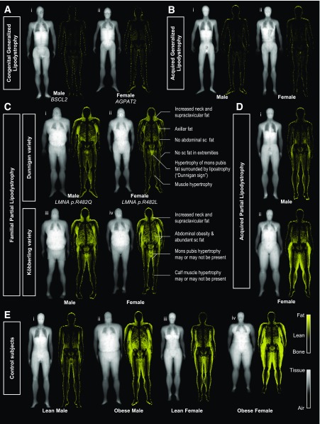
Research design and methods: We developed a new method from DXA scans called a "fat shadow," which is a color-coded representation highlighting only the fat tissue. We conducted a blinded retrospective validation study to assess its usefulness for the diagnosis of lipodystrophy syndromes.

Results: We evaluated the fat shadows from 16 patients (11 female and 5 male) with generalized lipodystrophy (GL), 57 (50 female and 7 male) with familial partial lipodystrophy (FPLD), 2 (1 female and 1 male) with acquired partial lipodystrophy, and 126 (90 female and 36 male) control subjects. FPLD was differentiated from control subjects with 85% sensitivity and 96% specificity (95% CIs 72-93 and 91-99, respectively). GL was differentiated from nonobese control subjects with 100% sensitivity and specificity (95% CIs 79-100 and 92-100, respectively).

Conclusions: Fat shadows provided sufficient qualitative information to infer clinical phenotype and differentiate these patients from appropriate control subjects. We propose that this method could be used to support the diagnosis.


Figures

Figure 1
Figure 1
Fat shadows derived from DXA scans of patients with lipodystrophy and selected control subjects. A: Patients with CGL have minimal fat signal on the fat shadow. CGL type 2 (i) was associated with no visible residual fat depots, whereas CGL type 1 (ii) is seen to have residual fat in the periauricular area and soles and in females the labial region. B: Male (i) and Female (ii) acquired GL patients are shown. They have no subcutaneous fat depots throughout their body, with minimal fat signal visible in the scan. C: The two classic presentations of different FPLD subtypes. FPLD2 (Dunnigan variety; i and ii) typically presents with the same signature phenotype with absence of subcutaneous fat in the extremities, hips, and abdomen (flanks), along with a hypertrophy in the neck, axillae, and mons pubis. Dunnigan sign was defined as the isolated high signal area corresponding to the mons pubis, surrounded by profound subcutaneous (sc) lipoatrophy. This sign was found to be helpful in identifying patients with FPLD2 and also evident in the one male with FPLD2 present in this cohort (i). Increased fat signal around the mons pubis region was present in all 21 (100%) FPLD2 females versus only 10 out of 29 (34%) FPLDX females and 0 out of 90 (0%) control females (95% CIs 84–100%, 18–54%, and 0–4%, respectively). FPLD1 (Köbberling variety; iii and iv) had neck hypertrophy and abdominal obesity. Calf muscle hypertrophy was present in 8 out of 35 patients with FPLDX (23%; 95% CIs 10–40). FPLDX population in its entirety was very heterogeneous, possibly due to heterogeneity in etiology (15). See Supplementary Fig. 4 and the Fat Shadow Atlas in the Supplementary Data for details. D: Acquired partial lipodystrophy: patients have acquired loss of fat from the upper half of the body with unknown cause. Fat depots in the lower half are spared and may be normal (i) or hypertrophied (ii). E: A selection of lean (i, iii) and obese (ii, iv) control subjects with sex-representative android/gynoid ratios and without metabolic disease. AGPAT2, 1-Acylglycerol-3-phosphate O-acyltransferase 2; BSCL2, Berardinelli-Seip congenital lipodystrophy 2; FPLDX, a broad group that includes FPLD1, FPLD3, and other types not numbered in standard nomenclature system; LMNA, lamin A/C gene.

References

    1. Capeau J, Magré J, Caron-Debarle M, et al. . Human lipodystrophies: genetic and acquired diseases of adipose tissue. In Adipose Tissue Development. Basel, Switzerland, Karger Publishers, 2010, p. 1–20 - PMC - PubMed
    1. Huang-Doran I, Sleigh A, Rochford JJ, O’Rahilly S, Savage DB. Lipodystrophy: metabolic insights from a rare disorder. J Endocrinol 2010;207:245–255 - PubMed
    1. Brown RJ, Araujo-Vilar D, Cheung PT, et al. . The diagnosis and management of lipodystrophy syndromes: a multi-society practice guideline. J Clin Endocrinol Metab 2016;101:4500–4511 - PMC - PubMed
    1. Guillín-Amarelle C, Sánchez-Iglesias S, Castro-Pais A, et al. . Type 1 familial partial lipodystrophy: understanding the Köbberling syndrome. Endocrine 2016;54:411–421 - PubMed
    1. Ajluni N, Meral R, Neidert AH, et al. . Spectrum of disease associated with partial lipodystrophy: lessons from a trial cohort. Clin Endocrinol (Oxf) 2017;86:698–707 - PMC - PubMed

Publication types