

doi: 10.3324/haematol.2018.196568.
Epub 2018 Sep 20.
Bone marrow mesenchymal stem/stromal cells from risk-stratified acute myeloid leukemia patients are anti-inflammatory in in vivo preclinical models of hematopoietic reconstitution and severe colitis
Affiliations
- PMID: 30237260
- PMCID: PMC6355484
- DOI: 10.3324/haematol.2018.196568
Item in Clipboard
Bone marrow mesenchymal stem/stromal cells from risk-stratified acute myeloid leukemia patients are anti-inflammatory in in vivo preclinical models of hematopoietic reconstitution and severe colitis
Haematologica.
2019 Feb.
No abstract available
Figures
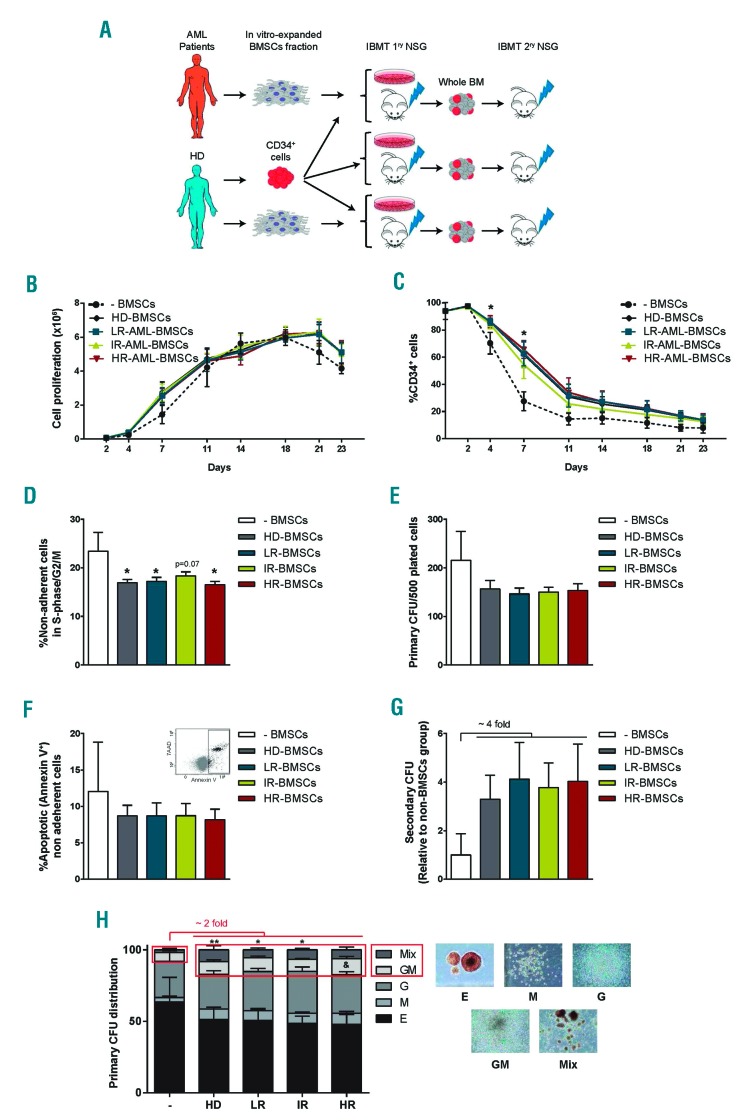
BMSCs from risk-stratified AML patients support in vitro homeostasis of CB-CD34+ HSPCs cells. (A) Schematic depicting the experimental design. (B) In vitro expansion kinetics of CB-CD34+ HSPCs alone (n=9) or co-cultured on BMSCs from HD (n=9) and AML patients (n=27; LR, n=9; IR, n=9; HR, n=9). This “n” is applicable to all subsequent graphs of this figure. (C) Kinetics of loss of CD34 antigen over time in CD34:BMSCs co-cultures. (D) Proportion of cycling CD34+ cells measured at day 11 of CD34:BMSCs co-cultures. (E) Proportion of apoptotic CB-CD34+ cells (Annexin V+) measured at day 11 of CD34:BMSCs co-cultures. (F) The primary clonogenic potential of CB-CD34+ HSPCs previously co-cultured 4 days with HD- or AML-BMSCs. (G) Clonogenic potential of CD34+ progenitors replated in secondary CFC assays normalized against -BMSCs. CFU-Mix (also known as CFU-GEMM) is a Colony-Forming Unit-Granulocyte, Erythrocyte, Monocyte/macrophage, Megakaryocyte. (H) Scoring of primary CFCs. CD34+ cells not previously exposed to BMSCs, were used as base-line control for CD34: BMSCs co-cultures. Because HSPCs gradually lose CD34 expression, we refer to them, we refer to them as “non-adherent cells” rather than “CD34+ cells”. *P< 0.05; ** P<0.01 for CFU-Mix and & P<0.05 for CFU-GM between BMSC groups and -BMSCs.
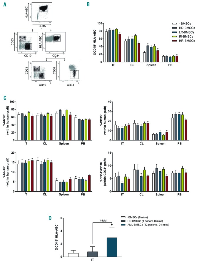
BMSCs from risk-stratified AML patients do not impair in vivo multilineage repopulating function of CB-CD34+ HSPCs. (A) Representative FACS analysis of the graft. The human graft is identified as the CD45+HLA-ABC+ fraction. The CD45+ human graft comprises B-lymphoid cells (CD19+), myeloid cells (CD33+) as well as immature CD34+ and CD34+CD38− cells. (B) Long-term multilineage hematopoietic reconstitution in the IT, CL, PB and spleen of NSG mice sacrificed 6-7 weeks after intra BM injection of CB-CD34+ HSPCs alone (n=5) or co-cultured for 4 days in BMSCs from HD (n=5) or AML patients (n=15; LR, n=5; IR, n=5; HR, n=5). n=90 mice, 4 per donor/patient. (C) Multilineage and multiorgan human chimerism demonstrating that AML-BMSCs do not negatively impact migration of HSPCs from the IT. No differences in the graft composition were found between CD34+ HSPCs co-cultured with BMSCs from HD or risk-stratified AML patients. (D) Long-term BM hematopoietic reconstitution assessed upon serial transplantation of primografted cells. IT: injected tibia; CL: contralateral tibia and femur; PB: peripheral blood.
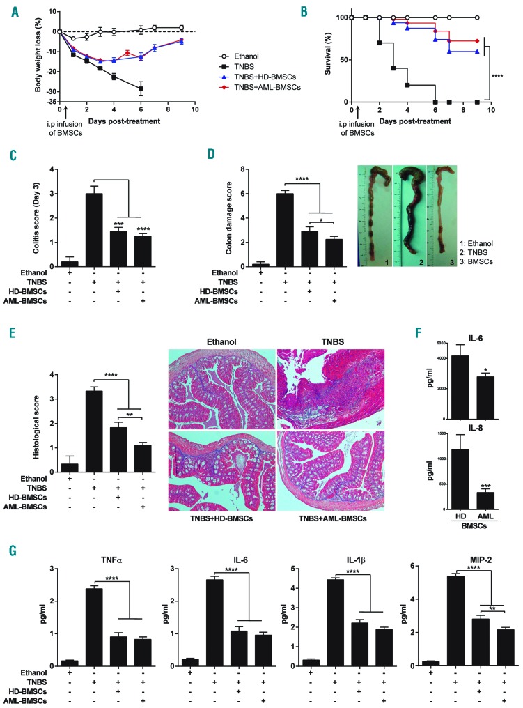
BMSCs from AML patients are highly anti-inflammatory in vivo in an experimental model of acute severe colitis. Colitis was induced by intracolonic administration of TNBS and mice were treated i.p. with PBS (TNBS group), HD-BMSCs or AML-BMSCs (106 cells) 12 hours after TNBS injection. Control mice received 50% ethanol. Clinical evolution was monitored by determining the daily body weight loss (A) as well as measuring the colitis score at day 3 (B), survival (C) the macroscopic colonic damage score (D), and the histopathologic score at day 3 (E). Data are expressed as mean±s.e.m. n=10 mice per group in a-d. n=5 mice per group in e. Statistical differences between groups were calculated as described in the methods section. (F) Serum levels on the indicated cytokines in colitis mice treated with either HD- or AML-BMSCs. (G) Levels of IL6 and IL8 produced by in vitro expanded HD- and AML-BMSCs. *P<0.05; **P<0.01; ***P<0.001; ****P<0.0001.
Similar articles
-
The use of unlicensed bone marrow-derived platelet lysate-expanded mesenchymal stromal cells in colitis: a pre-clinical study.Cytotherapy. 2019 Feb;21(2):175-188. doi: 10.1016/j.jcyt.2018.11.011. Epub 2019 Jan 2. Cytotherapy. 2019. PMID: 30611671
-
Mesenchymal Stromal Cells in Pediatric Hematopoietic Cell Transplantation a Review and a Pilot Study in Children Treated With Decidua Stromal Cells for Acute Graft-versus-Host Disease.Front Immunol. 2020 Oct 19;11:567210. doi: 10.3389/fimmu.2020.567210. eCollection 2020. Front Immunol. 2020. PMID: 33193339 Free PMC article.
-
Bone marrow stromal cell therapy improves survival after radiation injury but does not restore endogenous hematopoiesis.Sci Rep. 2020 Dec 17;10(1):22211. doi: 10.1038/s41598-020-79278-y. Sci Rep. 2020. PMID: 33335275 Free PMC article.
-
Significance of Cellular Cross-Talk in Stromal Vascular Fraction of Adipose Tissue in Neovascularization.Arterioscler Thromb Vasc Biol. 2019 Jun;39(6):1034-1044. doi: 10.1161/ATVBAHA.119.312425. Arterioscler Thromb Vasc Biol. 2019. PMID: 31018663 Free PMC article. Review.
-
MSC-Derived Extracellular Vesicles: New Emergency Treatment to Limit the Development of Radiation-Induced Hematopoietic Syndrome?Health Phys. 2020 Jul;119(1):21-36. doi: 10.1097/HP.0000000000001264. Health Phys. 2020. PMID: 32384375 Review.
Cited by
-
Robust In Vitro and In Vivo Immunosuppressive and Anti-inflammatory Properties of Inducible Caspase-9-mediated Apoptotic Mesenchymal Stromal/Stem Cell.Stem Cells Transl Med. 2022 Mar 3;11(1):88-96. doi: 10.1093/stcltm/szab007. Stem Cells Transl Med. 2022. PMID: 35641173 Free PMC article.
-
Engraftment characterization of risk-stratified AML in NSGS mice.Blood Adv. 2021 Dec 14;5(23):4842-4854. doi: 10.1182/bloodadvances.2020003958. Blood Adv. 2021. PMID: 34470043 Free PMC article.
-
High-efficient generation of VCAM-1+ mesenchymal stem cells with multidimensional superiorities in signatures and efficacy on aplastic anaemia mice.Cell Prolif. 2020 Aug;53(8):e12862. doi: 10.1111/cpr.12862. Epub 2020 Jun 29. Cell Prolif. 2020. PMID: 32597552 Free PMC article.
-
The Multi-Kinase Inhibitor EC-70124 Is a Promising Candidate for the Treatment of FLT3-ITD-Positive Acute Myeloid Leukemia.Cancers (Basel). 2022 Mar 21;14(6):1593. doi: 10.3390/cancers14061593. Cancers (Basel). 2022. PMID: 35326743 Free PMC article.
-
Effects of Resveratrol, Curcumin and Quercetin Supplementation on Bone Metabolism-A Systematic Review.Nutrients. 2022 Aug 26;14(17):3519. doi: 10.3390/nu14173519. Nutrients. 2022. PMID: 36079777 Free PMC article.
References
-
- Grimwade D, Lo Coco F. Acute promyelocytic leukemia: a model for the role of molecular diagnosis and residual disease monitoring in directing treatment approach in acute myeloid leukemia. Leukemia. 2002;16(10):1959–1973. - PubMed
Publication types
MeSH terms
Grants and funding
LinkOut - more resources
Full Text Sources
Other Literature Sources
