. 2018 Oct 2;115(40):E9288-E9297.
doi: 10.1073/pnas.1801759115. Epub 2018 Sep 20.

Synchronized mechanical oscillations at the cell-matrix interface in the formation of tensile tissue

Affiliations

Synchronized mechanical oscillations at the cell-matrix interface in the formation of tensile tissue

David F Holmes et al. Proc Natl Acad Sci U S A. .

Abstract

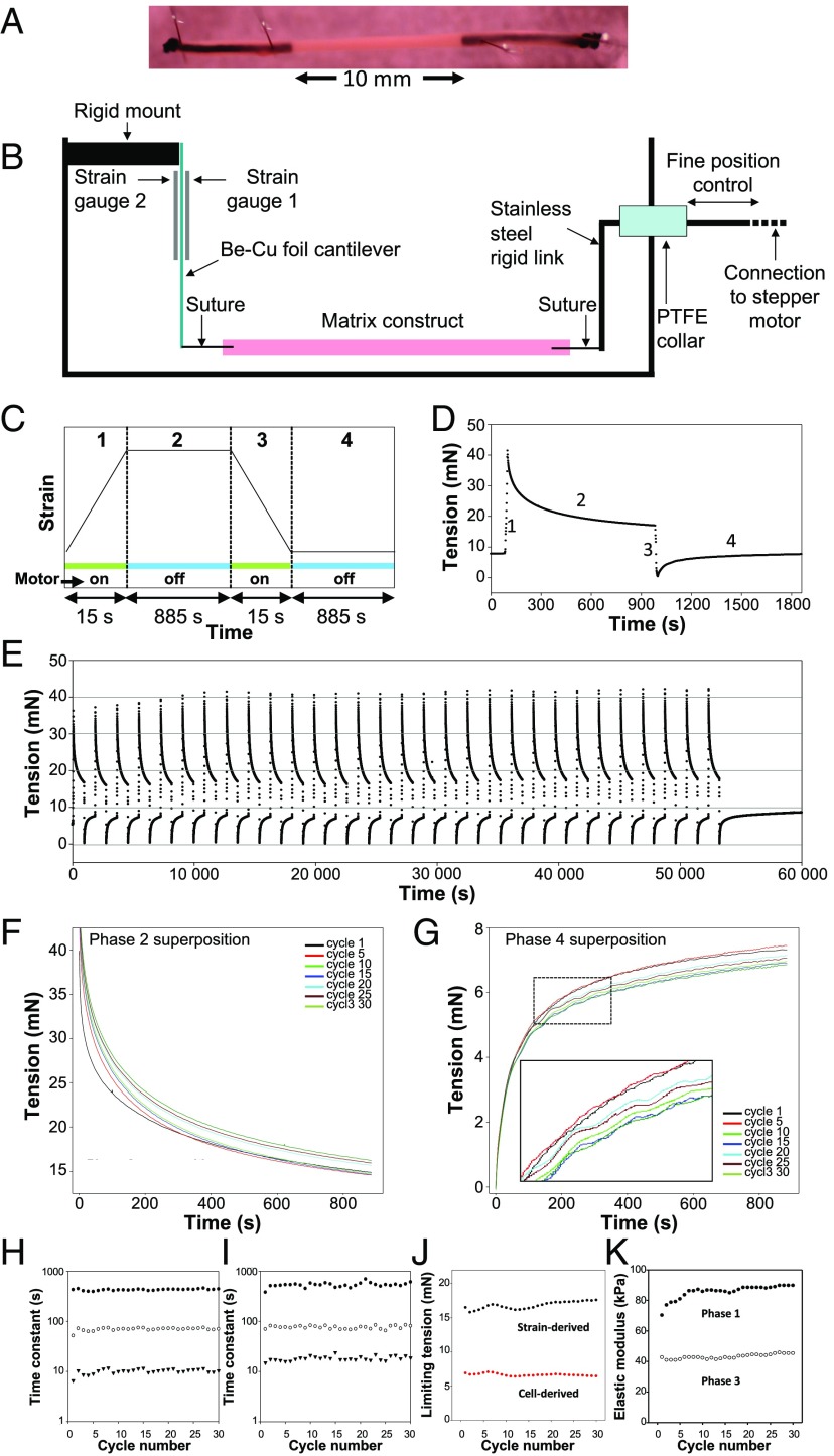
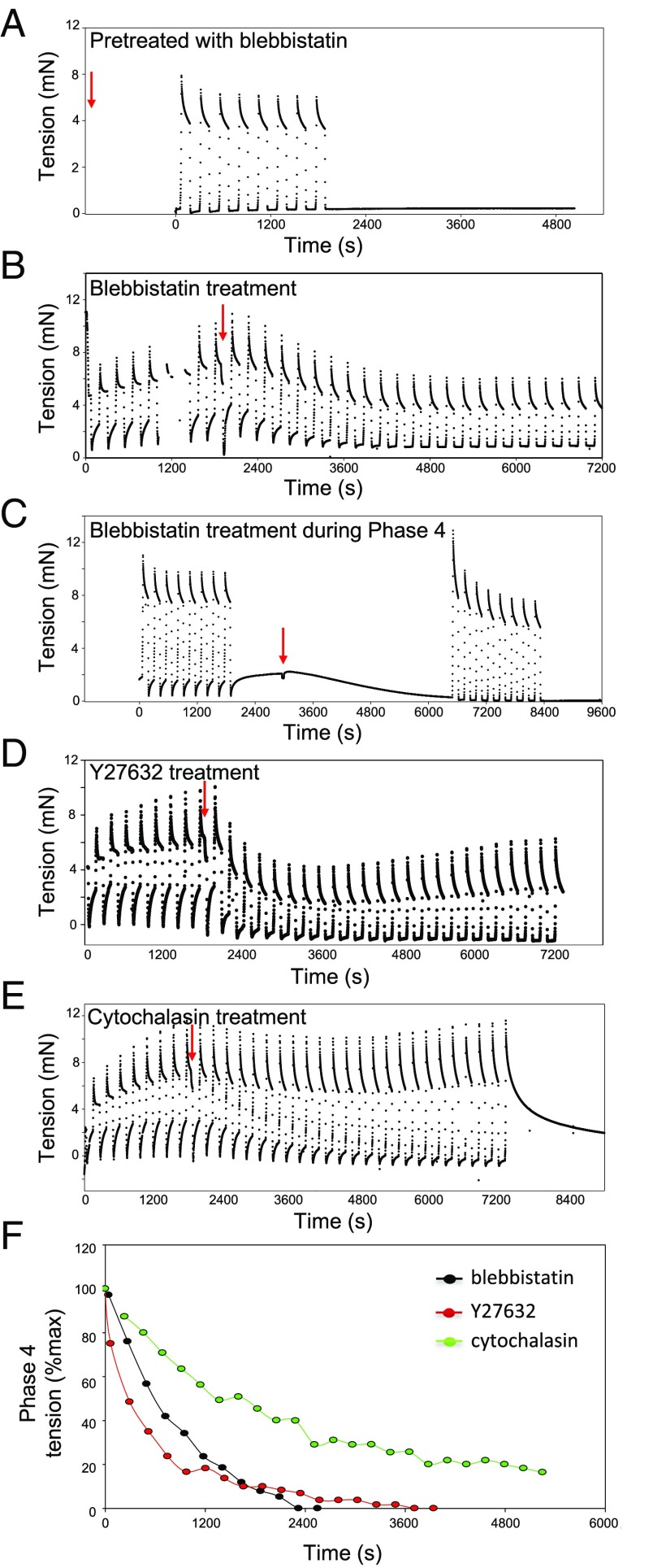
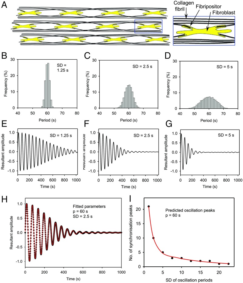
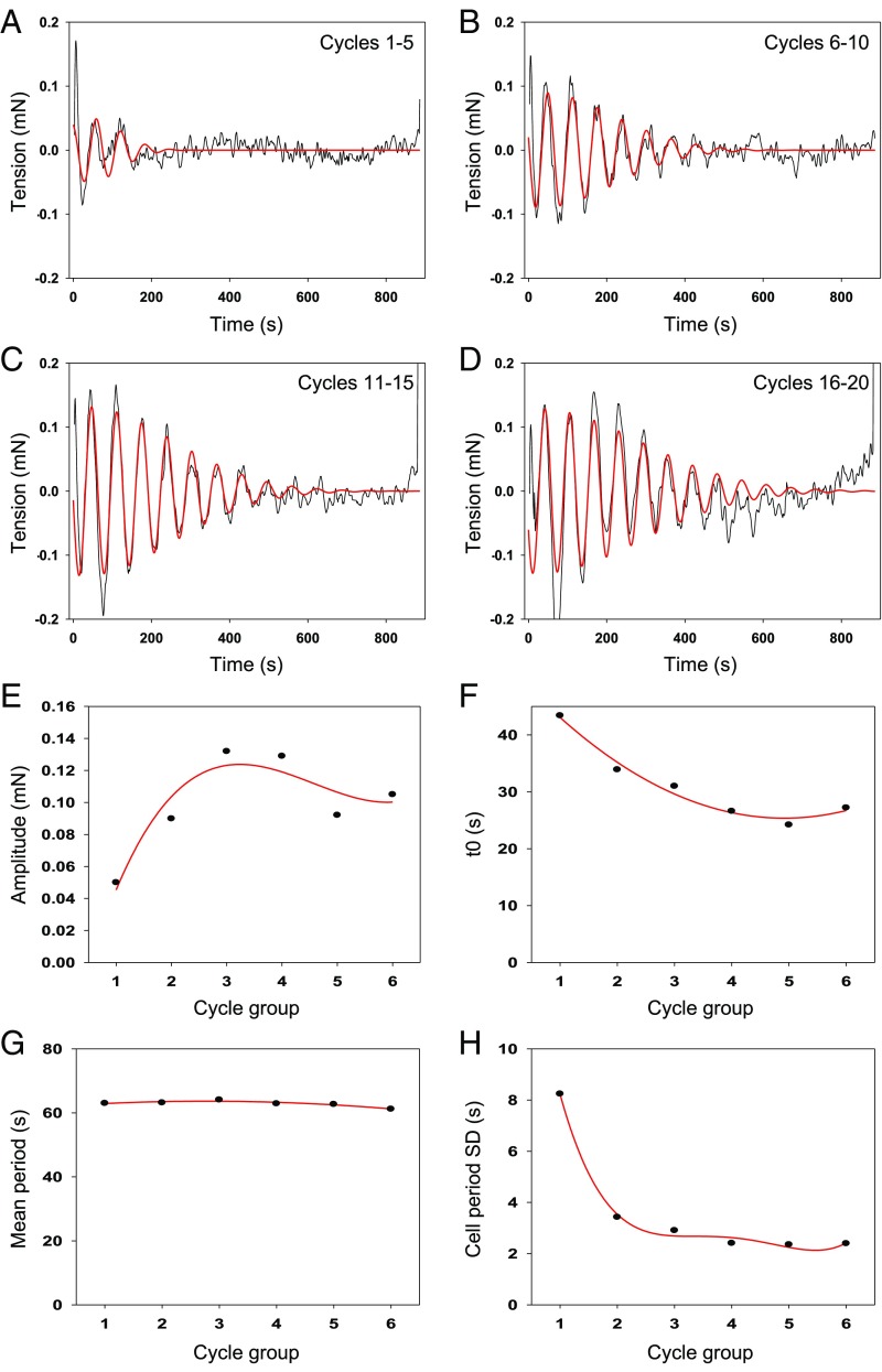
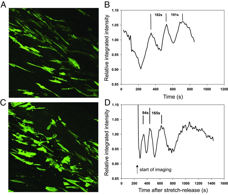
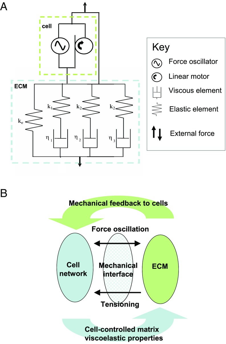
The formation of uniaxial fibrous tissues with defined viscoelastic properties implies the existence of an orchestrated mechanical interaction between the cytoskeleton and the extracellular matrix. This study addresses the nature of this interaction. The hypothesis is that this mechanical interplay underpins the mechanical development of the tissue. In embryonic tendon tissue, an early event in the development of a mechanically robust tissue is the interaction of the pointed tips of extracellular collagen fibrils with the fibroblast plasma membrane to form stable interface structures (fibripositors). Here, we used a fibroblast-generated tissue that is structurally and mechanically matched to embryonic tendon to demonstrate homeostasis of cell-derived and external strain-derived tension over repeated cycles of strain and relaxation. A cell-derived oscillatory tension component is evident in this matrix construct. This oscillatory tension involves synchronization of individual cell forces across the construct and is induced in each strain cycle by transient relaxation and transient tensioning of the tissue. The cell-derived tension along with the oscillatory component is absent in the presence of blebbistatin, which disrupts actinomyosin force generation of the cell. The time period of this oscillation (60-90 s) is well-defined in each tissue sample and matches a primary viscoelastic relaxation time. We hypothesize that this mechanical oscillation of fibroblasts with plasma membrane anchored collagen fibrils is a key factor in mechanical sensing and feedback regulation in the formation of tensile tissues.

Keywords: cell force; collagen; electron microscopy; fibroblast; tension.


Conflict of interest statement

The authors declare no conflict of interest.

Figures

Fig. 1.
Fig. 1.
Tension measurements on a cell–matrix construct during repeated strain cycles. (A) Image of typical matrix construct before loading into the tension stepper. (B) Schematic diagram to show the primary components of a modified well in a six-well plate. The construct is attached on the left-hand side to a beryllium–copper cantilever which has a strain gauge bonded to each surface. Electrical resistance changes in these strain gauges are used to measure the tension in the specimen. The other end of the construct is attached to a sliding rod, which is linked to a computer controlled linear actuator. (C) The standard strain cycle of four phases (labeled 1–4). The green bars indicate uniform strain rate (motor on) and blue bars constant strain (motor off). (D) Temporal change in the tension of the construct over one strain cycle showing the four characteristic phases. Phases 2 and 4 correspond to stress relaxation and tension recovery, respectively. (E) Continuous tension plot over 30 identical strain cycles, each of 30 min. (F) Superposition of a sample of tension curves from phase 2 extracted from the plot shown in E. (G) Corresponding superposition for phase 4. (H) Plot of the exponential time constants in phase 2 against cycle number. (I) Plot of the exponential fitted time constants in phase 4 against cycle number. (J) Plot of the limiting tension values in phases 2 (black circles) and 4 (red circles) plotted against cycle number. (K) Mean elastic modulus from phases 1 (closed circles) and 3 (open circles).
Fig. 2.
Fig. 2.
Inhibition of cell-derived tension. (A) Tension plot after pretreatment of the construct with blebbistatin. The tension recovery in phase 4 is abolished. (B) Tension plot showing the effect of adding the blebbistatin after eight strain cycles. The tension established in each successive phase 4 decays over ∼40 min and remains blocked in the subsequent strain cycles. (C) Addition of blebbistatin to a static construct in late phase 4 leads to a loss of tension over ∼60 s. Tension recovery is subsequently blocked in continued strain cycles. (D) ROCK inhibitor also abolishes the tension recovery phase over a similar time period to the blebbistatin treatment. (E) Addition of cytochalasin progressively reduces the tension recovery over ∼60 min, but ∼20% of the original tension recovery shows a slower decay. (F) Plots of tension recovery level vs. time after addition of inhibitor.
Fig. 3.
Fig. 3.
Synchronization model of cell-derived tension oscillation: simulations of a transiently synchronized set of 104 cellular mechanical oscillators. (A) Schematic depiction of the mechanical connectivity of the cell–matrix construct. The cells (shown in yellow) are embedded within an aligned network of collagen fibrils (shown as black lines). The fibripositor structures containing individual collagen fibrils are shown as mechanical connections between the cytoskeleton and the extracellular collagen fibrillar matrix. (BD) Gaussian distributions of oscillation period with mean period 60 s and (B) SD = 1.25 s, (C) SD = 2.5 s, and (D) SD = 5 s. The corresponding resultant forces after transient synchronization (t = 0) are shown in EG, respectively. For Gaussian distribution of oscillation period and cell number >∼1,000, the summed force oscillation can be represented analytically by Eq. 3. (H) The P = 60, SD = 2.5 simulation (solid circles) fitted using linear least squares to this decaying oscillation function (solid line). The predicted number of oscillation peaks for P = 60 is shown in I as a function of the SD of the oscillation period.
Fig. 4.
Fig. 4.
Fitting of the synchronization model to cell-derived tension oscillation. (AD) Experimental plots of residual tension after three-exponential fitting vs. time for cell-derived tension recovery. The residual data have been averaged over five successive cycles for six groups covering 30 strain cycles. The average tension plots are fitted to the exponentially damped sinusoid from the oscillation model (red lines). The examples shown are (A) cycles 1–5, (B) cycles 6–10, (C) cycles 11–15, and (D) 16–20. In each case the data were fitted to the model analytical function to yield values of amplitude (E), time offset (F), mean period (G), and SD of the cell period distribution (H).
Fig. 5.
Fig. 5.
Confocal microscope imaging of cellular oscillation. (A and B) Undisturbed static construct. The plot shows the integrated intensity over a single cell during a time-lapse series; an oscillation with a period ∼3 min was observed. (C and D) Data similar to A and B on a construct after ∼10% strain for ∼60 s and subsequent release to the original length. Imaging starts ∼90 s after release and shows a reduced period oscillation (∼90 s) which progressively relaxes toward that of the unstrained constructs.
Fig. 6.
Fig. 6.
Models relating the mechanical interplay between cells and the ECM construct. (A) A simple combination of mechanical elements that generates the key observed mechanical behavior of the cell–matrix construct. The parameters k1, k2, and k3 are the elastic constants; the parameters η1, η2, and η3 are the viscous constants. (B) A simple working model of cell–matrix interaction where cells tension the resting matrix, receive mechanical feedback signals, and can maintain a matrix of defined viscoelastic properties.

Similar articles

Cited by

References

    1. Lee S, Kumar S. Actomyosin stress fiber mechanosensing in 2D and 3D. F1000 Res. 2016;5:F1000. - PMC - PubMed
    1. Labouesse M. Role of the extracellular matrix in epithelial morphogenesis: A view from C. elegans. Organogenesis. 2012;8:65–70. - PMC - PubMed
    1. Engler AJ, Sen S, Sweeney HL, Discher DE. Matrix elasticity directs stem cell lineage specification. Cell. 2006;126:677–689. - PubMed
    1. Cameron AR, Frith JE, Cooper-White JJ. The influence of substrate creep on mesenchymal stem cell behaviour and phenotype. Biomaterials. 2011;32:5979–5993. - PubMed
    1. Cameron AR, Frith JE, Gomez GA, Yap AS, Cooper-White JJ. The effect of time-dependent deformation of viscoelastic hydrogels on myogenic induction and Rac1 activity in mesenchymal stem cells. Biomaterials. 2014;35:1857–1868. - PubMed

Publication types