. 2018 Sep 21;361(6408):eaat5236.
doi: 10.1126/science.aat5236.

A gut-brain neural circuit for nutrient sensory transduction

Affiliations

A gut-brain neural circuit for nutrient sensory transduction

Melanie Maya Kaelberer et al. Science. .

Abstract

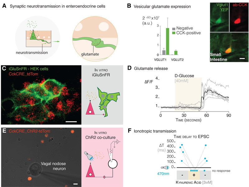
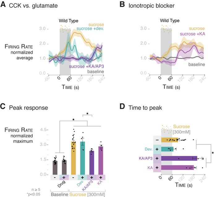
The brain is thought to sense gut stimuli only via the passive release of hormones. This is because no connection has been described between the vagus and the putative gut epithelial sensor cell-the enteroendocrine cell. However, these electrically excitable cells contain several features of epithelial transducers. Using a mouse model, we found that enteroendocrine cells synapse with vagal neurons to transduce gut luminal signals in milliseconds by using glutamate as a neurotransmitter. These synaptically connected enteroendocrine cells are referred to henceforth as neuropod cells. The neuroepithelial circuit they form connects the intestinal lumen to the brainstem in one synapse, opening a physical conduit for the brain to sense gut stimuli with the temporal precision and topographical resolution of a synapse.


Conflict of interest statement

Competing interests: Some of the findings in this manuscript have been used to file a provisional patent application. No other competing interests are declared.

Figures

Fig. 1.
Fig. 1.. Enteroendocrine cells contact sensory nerve fibers.
(A) CckGFP_Pgp9.5GFP mice express GFP in CCK-enteroendocrine cells and Pgp9.5 sensory nerve fibers. The two cell types are shown in the enlarged view, with the CCK- enteroendocrine cell represented by a triangle. (B) Confocal microscopy image of proximal small intestine villus showing a GFP-labeled CCK-enteroendocrine cell and GFP-labeled Pgp9.5 nerve fibers; 18.9 ± 2.0% SEM of CckGFP cells contact Pgp9.5 fibers (n = 3 mice, >100 cells per mouse). (C) PYY-stained enteroendocrine cells (left, green) in the colon contact Phox2b vagal nerve fibers (center, red) in a Phox2bCRE_tdTomato mouse; merged image is shown on the right. (D) Two-thirds of CckGFP (green) enteroendocrine cells colocalize with the presynaptic marker synapsin-1 (purple) (n = 6 mice, 200 cells per mouse). (E) Real-time quantitative polymerase chain reaction (qPCR) expression levels of presynaptic transcripts, including genes encoding for synaptic adhesion proteins (n = 3 mice, >10,000 cells per cell type per mouse; error bars indicate mean ± SEM; a.u., arbitrary units; EEC, enteroendocrine cell). All scale bars, 10 μm.
Fig. 2.
Fig. 2.. Enteroendocrine cells of the colon and small intestine synapse with vagal nodose neurons.
(A) Model of AG-rabies-GFP enema delivery. (B) PYY cells expressing tdTomato (top left, red) are infected by ΔG-rabies-GFP (top right, green). Overlay (bottom) shows overlap of 87.8 ± 2.4% SEM (n = 5 mice). In the absence of G glycoprotein (ΔG), AG-rabies-GFP does not spread beyond the infected PYY cell. (C) EnvA-ΔG-rabies-GFP virus enters cells via the TvA receptor and spreads by using the rabG protein within specific cells. (D) EnvA-ΔG-rabies-GFP (top right, green) infects PYY cells (top left, red) and spreads synaptically to underlying colon nerve fibers. Three-dimensional reconstruction (bottom) shows EnvA-ΔG-rabies-GFP-infected PYY cell and mono- synaptically labeled nerve fiber. (E) EnvA-ΔG-rabies-GFP enema infects colonic enteroendocrine cells and spreads onto vagal neurons in the nodose ganglion (green). (F) In additional experiments, ΔG-rabies-GFP delivered by oral gavage spreads in the intestinal lumen of CckCRE_rabG-TvA mice to label the nucleus tractus solitarius (green). This neuroepithelial circuit links the intestinal lumen with the brainstem. The inset shows the location of the nucleus tractus solitarius in the mouse brain. All scale bars, 10 μm.
Fig. 3.
Fig. 3.. Enteroendocrine cells transduce glucose stimuli onto vagal neurons.
(A) Model of intestinal intraluminal perfusion and vagal nerve electrophysiology. (B) Normalized traces for baseline, Ensure, 300 mM sucrose, and 300 mM sucrose with 3 mM phloridzin (phl) in wild-type mice. Gray bar indicates treatment period; shading indicates SEM. (C) Ensure, 300 mM sucrose, and 150 mM D-glucose stimulate vagal firing rate, which is abolished by SGLT1-blocker phloridzin [n ≥ 5 mice; *P < 0.0001, analysis of variance (ANOVA) with post hocTukey’s HSD test; error bars indicate SEM]. (D) Intestinal epithelial cells express Sgltl, but nodose neurons do not (n = 3 mice, >10,000 cells per cell type per mouse; data are presented as mean ± SEM). (E) Nodose neurons cultured alone for electrophysiology (widefield microscopy image on left, model on right). (F) Nodose neurons do not respond to 10 mM glucose in voltage-clamp (left trace) or current-clamp (right trace) mode. Insets show that neurons respond to voltage or current pulse, indicating viability. (G) Nodose neurons cocultured with GFP-positive enteroendocrine cells for electrophysiology (image on left, model on right). Innervated enteroendocrine cells are shown at the bottom. (H) In coculture, glucose evoked EPSCs (top left) and action potentials (top right) in connected neurons (scale of current or voltage and time are shown below the traces). Dashed-line box indicates action potentials expanded in right inset. Quantification of EPSC amplitude and frequency (bottom left and center; n = 21 neurons alone; n = 6 neurons connected to enteroendocrine cells) and action potentials (bottom right; n = 21 alone; n = 5 neurons connected to enteroendocrine cells) in GFP-negative (−) and -positive (+) cells. All scale bars, 10 μm.
Fig. 4.
Fig. 4.. Millisecond transduction from enteroendocrine cells to vagal neurons.
(A) Model of intraluminal photostimulation and vagal electrophysiology. (B) In CckCRE_ChR2-tdTomato mice, intestinal enteroendocrine cells express ChR2. (C) Normalized traces for 473-nm intraluminal laser, 300 mM sucrose, and baseline in CckCRE_ChR2 mice. Shading indicates SEM. (D) 473-nm intraluminal laser stimulates vagal firing rate in CckCRE_ChR2, but not wild-type, mice (n ≥ 5 mice; *P < 0.05, ANOVA with post hocTukey’s HSD test; error bars indicate SEM). (E) Patch-clamp electrophysiology of neurons (model on left) in coculture with CckCRE_ChR2 cells (image on right). (F) In coculture, 473-nm photostimulation evoked EPSCs (trace on left) in connected nodose neurons (quantification on right) (n = 9 neurons connected to enteroendocrine cells; -, neurons alone; +, neurons cocultured with enteroendocrine cells; ΔT, time between stimulus and onset of EPSCs). Scale of current and time is shown below the trace. (G) Model of intraluminal photoinhibition and vagal electrophysiology. (H) In CckCRE_Halo-YFP mice, intestinal enteroendocrine cells express halorhodopsin (eNpHR3.0). (I) Normalized traces for baseline, 300 mM sucrose, and 300 mM sucrose with 532-nm intraluminal laser. Shading indicates SEM. (J) In CckCRE_Halo, but not wild-type, mice, a 532-nm intraluminal laser abolishes the effect of sucrose on vagal firing rate (n ≥ 5 mice per group; *P <0.0001, ANOVA with post hocTukey’s HSD test; error bars indicate SEM). All scale bars, 10 μm.
Fig. 5.
Fig. 5.. Glutamate is used as a neurotransmitter between enteroendo- crine cells and neurons.
(A) Model of synaptic neurotransmission in enteroendocrine cells. (B) Enteroendocrine cells express the vesicular glutamate genes encoding VGLUT1 and 2 (Slc17a7 and Slc17a6) (quantification by qPCR on left, confocal microscopy images on right). (C) CckCRE_tdTomato enteroendocrine cells were cocultured with HEK cells that express the glutamate sniffer protein, iGluSnFR (multiphoton microscopy image on left, model on right). (D) A stimulus of 40 mM D-glucose administered during the time period indicated by the beige shading elicits a response in iGluSnFR-HEK cells (n = 3 cultures; individual cell, gray trace; average of all cells, black trace). ΔF/F, difference in fluorescence intensity between resting state and after stimulus. (E) Coculture with neurons and CckCRE_ChR2 cells (multiphoton microscopy image on left) for electrophysiology of neurons and microperfusion of the glutamate-receptor blocker kynurenic acid (model on right). (F) In coculture, 473-nm photostimulation evoked EPSCs in connected nodose neurons, these currents were abolished, and no response was observed with the addition (+) of 3 mM kynurenic acid. The response was recovered after the drug was washed off (indicated by second “-” condition on right) (n = 4 neurons connected to enteroendocrine cells). All scale bars, 10 μm.
Fig. 6.
Fig. 6.. The rapid vagal response to sucrose is dependent on glutamate, whereas CCK contributes to the prolonged response.
(A) Normalized traces for baseline, 300 mM sucrose, 300 mM sucrose after treatment with 2 mg/kg devazepide, and 300 mM sucrose after treatment with glutamate inhibitor cocktail KA/AP3 [150 mg/kg kynurenic acid (KA) with 1 mg/kg DL-2-amino-3-phosphonoproprionic acid (AP-3)] in wild-type mice. Shading indicates SEM. (B) Normalized traces for baseline, 300 mM sucrose, and 300 mM sucrose after treatment with 150 mg/kg KA in wild-type mice. Shading indicates SEM. (C) KA/AP3 attenuates the maximum normalized vagal firing rate in response to sucrose, whereas devazepide and KA alone do not. (D) KA/AP3 and KA alone prolong the time to peak from an average of 92.8 s to 198 and 179 s, respectively. Devazepide (2 mg/kg) does not significantly change the time to peak (mean = 67.1 s). For (C) and (D), n ≥ 5 mice per group; *P < 0.05, ANOVA with post hocTukey’s HSD test; error bars indicate SEM.

Comment in

References

    1. Alberts B, Bray D, Lewis J, Raff M, Roberts K, Watson JD, Molecular Biology of the Cell (Garland, ed. 3, 1994), pp. 907–982.
    1. Psichas A, Reimann F, Gribble FM, Gut chemosensing mechanisms. J. Clin. Invest 125, 908–917 (2015). doi: 10.1172/JCI76309; pmid: - DOI - PMC - PubMed
    1. Furness JB, Rivera LR, Cho HJ, Bravo DM, Callaghan B, The gut as a sensory organ. Nat. Rev. Gastroenterol. Hepatol 10, 729–740 (2013). doi: 10.1038/nrgastro.2013.180; pmid: - DOI - PubMed
    1. Feyrter F, Über diffuse endokrine epitheliale Organe (J. A. Barth, Leipzig, Germany, 1938).
    1. Rehfeld JF, The new biology of gastrointestinal hormones. Physiol. Rev 78, 1087–1108 (1998). doi: 10.1152/physrev.1998.78.4.1087; pmid: - DOI - PubMed

Publication types

Substances