. 2019 Feb;38(7):1136-1150.
doi: 10.1038/s41388-018-0501-z. Epub 2018 Sep 20.

A reciprocal feedback between the PDZ binding kinase and androgen receptor drives prostate cancer

Affiliations

A reciprocal feedback between the PDZ binding kinase and androgen receptor drives prostate cancer

Anne Y Warren et al. Oncogene. 2019 Feb.

Abstract

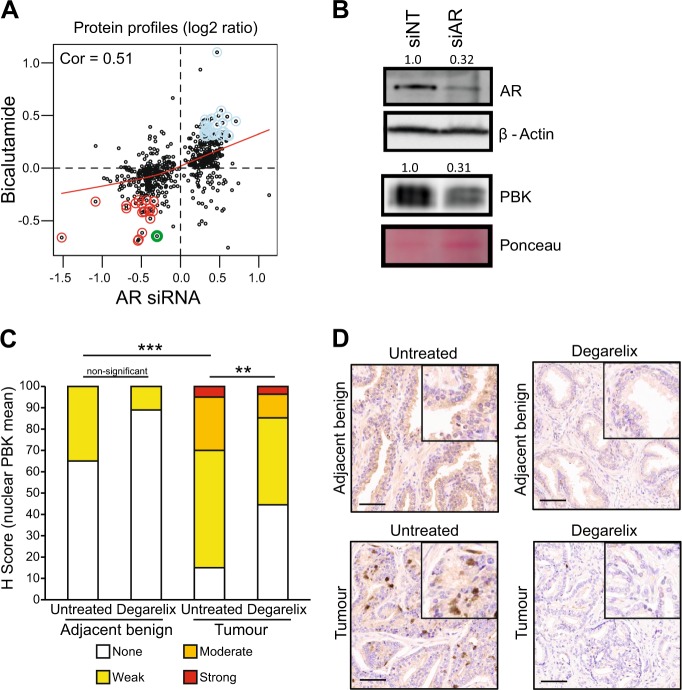
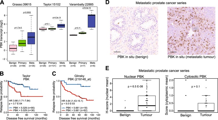
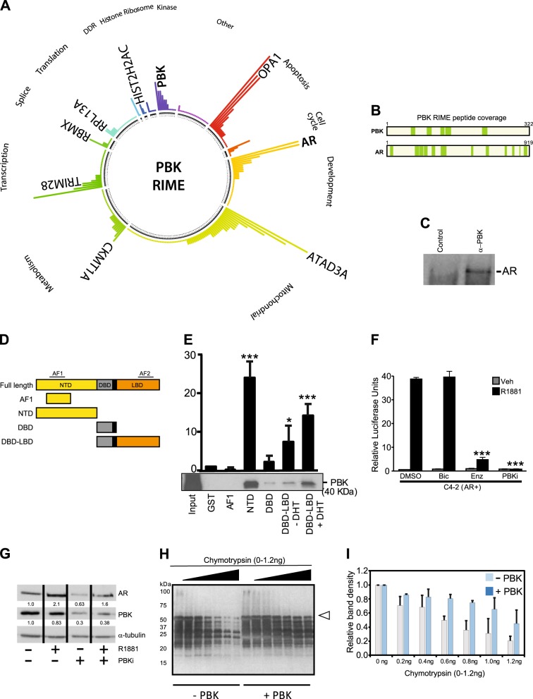
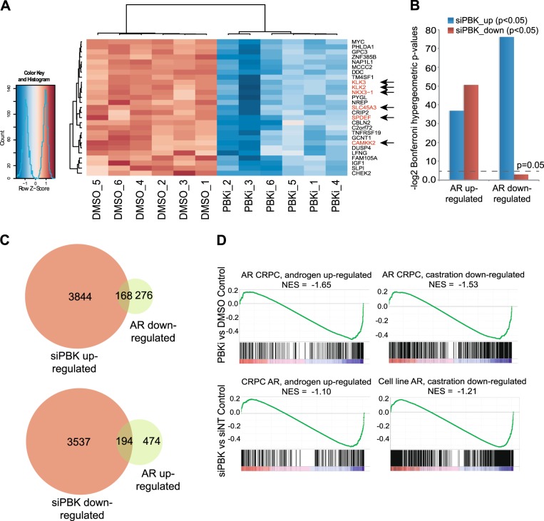
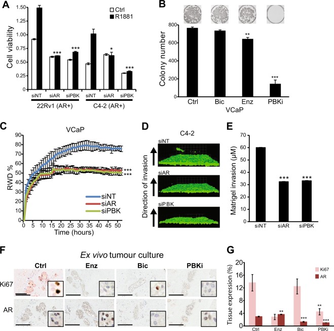
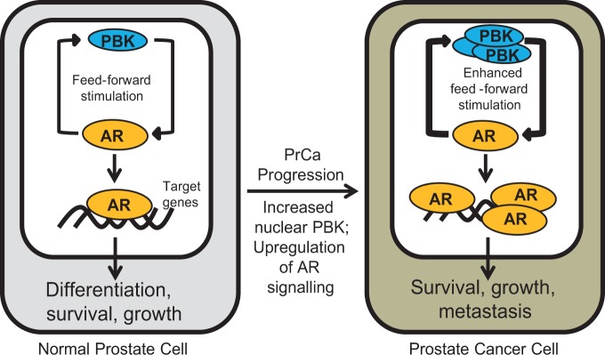
Elucidation of mechanisms underlying the increased androgen receptor (AR) activity and subsequent development of aggressive prostate cancer (PrCa) is pivotal in developing new therapies. Using a systems biology approach, we interrogated the AR-regulated proteome and identified PDZ binding kinase (PBK) as a novel AR-regulated protein that regulates full-length AR and AR variants (ARVs) activity in PrCa. PBK overexpression in aggressive PrCa is associated with early biochemical relapse and poor clinical outcome. In addition to its carboxy terminus ligand-binding domain, PBK directly interacts with the amino terminus transactivation domain of the AR to stabilise it thereby leading to increased AR protein expression observed in PrCa. Transcriptome sequencing revealed that PBK is a mediator of global AR signalling with key roles in regulating tumour invasion and metastasis. PBK inhibition decreased growth of PrCa cell lines and clinical specimen cultured ex vivo. We uncovered a novel interplay between AR and PBK that results in increased AR and ARVs expression that executes AR-mediated growth and progression of PrCa, with implications for the development of PBK inhibitors for the treatment of aggressive PrCa.


Conflict of interest statement

The authors declare that they have no conflict of interest.

Figures

Fig. 1
Fig. 1
Identification of androgen-stabilised proteome in prostate cancer (PrCa). a Scatterplot showing the relationship between protein levels following AR knockdown and AR antagonist treatment of C4-2 cells grown in full media. Red line shows lowess regression, red points show commonly downregulated proteins, light blue points show commonly upregulated proteins (cor = 0.51). PBK is shown in green. b Western blot analysis showing of expression of PBK and AR proteins. C4-2 cells were transiently transfected with small interfering (si)-RNA targeting AR (siAR) or with non-targeting control siRNA (siNT); β-actin is the loading control for AR and the Ponceau for PBK. c Immunohistochemical staining score (H score) of nuclear PBK protein expression in tumours (in triplicates) and untreated benign adjacent epithelia (in duplicates) in patients with (n = 27) or without (n = 20) 7 days’ treatment with the LHRH analogue degarelix, statistical significance calculated by Mann–Whitney test. d Representative IHC images of corresponding tissue samples from degarelix-treated and untreated patients. Scale bars = 50 µm. H score ranged from 0 to 9 and converted it to a 4-point scale as 0 = none, 1–3 = weak; 4–6 = moderate and 7–9 = strong
Fig. 2
Fig. 2
Clinical relevance of PDZ binding kinase (PBK) in PrCa. a Boxplots show PBK transcript expression in prostate tissues in three independent gene expression data sets [–22]. Gene expression values (log2) of PBK trancript are shown. bc Kaplan–Meier survival curve from recursive partitioning analysis. High levels of PBK transcript levels are associated with poor recurrence-free survival in the b Taylor cohort [21] and c in Glinsky data set [23]. d Representative IHC images of prostate samples from benign prostate glands and hormone refractory (metastatic) tumour tissue. Scale bars = 50 µm. ef Corresponding Boxplot showing e nuclear and f cytosolic PBK protein levels in metastatic tumours from PrCa patients (n = 50), and benign specimen (n = 50) (p < 6.46e-06; Wilcoxon test for IHC)
Fig. 3
Fig. 3
Interaction of PBK with AR and its effect on AR stability. a PBK RIME summary plot showing gene ontology categories (DAVID GO analysis) and key PBK-associated proteins identified in this experiment (filtered using IgG RIME) in C4-2 cells. Bar heights represent the unique peptide counts for each protein identified and the figure was drawn using the ‘circlize’ library in the R statistical package. b Peptide coverage of the PBK and AR proteins following PBK RIME assay in C4-2 cells. The locations of the identified peptides are highlighted in green blocks. c Co-immunoprecipitation showing interaction of endogenous AR with PBK. C4-2 Cell lysate were incubated with the PBK antibody followed by Western blot analysis to detect AR protein. A representative blot is shown (n = 3). d–e GST pull-down assay; d Schematic representation of AR domains used in pull-down. e GST alone and GST-tagged AR domains were incubated with recombinant PBK for 2 h and subjected to a pull-down assay. PBK was detected by immunoblotting with an anti-PBK antibody. The AR-DBD-LBD was expressed, purified and incubated ± 50 μM DHT. Fold interaction of each domain with PBK based on the intensity of the band as calculated by Image J software, GST = 1. A representative blot is shown (n = 2–5). f Luciferase reporter assay. AR transactivation potential in C4-2 cells transiently transfected with MMTV-luciferase plasmid and treated with androgen (1 nM R1881), bicalutamide, enzalutamide or PBKi (all 10 µM); (p < 0.001) bars show mean ± SD (n = 3). p values for two-sided Student’s t test. *** = p < 0.001. g Western blot showing protein expression of PBK and AR in C4-2 cells. Cells were treated with R1881 (1 nM) or PBKi (1 µM) for 48 h; Tubulin is the loading control. A representative blot is shown (n = 3). h Representative Western blot for ARNTD digested with chymotrypsin in the absence or presence of recombinant PBK. Open triangle represents the full-length ARNTD. Proteolysis was performed for 5 min in the presence of increasing concentrations of chymotrypsin (0–1.2 ng). ARNTD fragments were detected by Western blot using the anti-androgen receptor antibody ab3510 (Abcam) corresponding to human AR amino acids 1–21 (N-terminal). i Quantitation of the ARNTD full-length polypeptide, after digestion with chymotrypsin for two independent experiments. PBK = PDZ binding kinase; AR = androgen receptor; IP = immunoprecipitation; AF = activation function; NTD = amino terminal domain; DBD = DNA binding domain; LBD = ligand-binding domain; GST = glutathione-s-transferase; DHT = dihydrotestosterone, DMSO = dimethylsulphoxide
Fig. 4
Fig. 4
The PBK-regulated transcriptional programme in PrCa. a Heatmap showing differentially expressed genes from RNAseq analysis of PBK inhibitor (PBKi) and vehicle control (DMSO) treated C4-2 prostate cancer cells. Heatmap shows z-scores and known AR-regulated genes are highlighted in red and with arrows. b Barplot showing significance testing of the overlaps between PBK inhibitor or PBK knockdown and AR gene sets. Bonferroni corrected p values from hypergeometric tests are plotted as inverse log-values. c Venn diagrams showing the overlap between PBK and AR-regulated genes (overlaps used to calculate p values in c. d Gene set enrichment analysis (GSEA) plots showing the enrichment of AR gene sets in the PBK inhibitor and PBK knockdown ranked expression profiles
Fig. 5
Fig. 5
Regulation of PrCa growth and invasion by PBK signalling. a MTS cell viability assay of 22Rυ1 and C4-2 cells transfected with indicated siRNA targeting AR (siAR) or PBK (siPBK) or non-targeting control (siNT). Cells were grown in the presence or absence of R1881 for 5 days or near confluence; bars show mean ± SD (n = 3). p values for two-sided Student’s t test. b Clonogenic cell survival assay in VCaP cell lines treated with bicalutamide, enzalutamide or PBKi (14-day assay, all at 10 µM). c Scratch–wound assay. VCaP cells were transiently transfection with siNT, siAR or siPBK and wound closure was monitored, error bars represent ± SD (n = 4). Significance calculated by two-way ANOVA. de Matrigel-based inverted invasion assay d photomicrographs and e quantification of GFP tagged C4-2 cells transiently transfected with indicated siRNA; bars show mean ± SD (n = 3), significance calculated by D’Agostino & Pearson omnibus normality test p < 0.0001. f Photomicrographs of PrCa tissue explants treated with indicated drugs. Scale bars = 200 µm. g Barplot shows nuclear expression of AR and Ki67 in human PrCa tissue grown ex vivo and treated with bicalutamide, enzalutamide or PBKi (10 µM); bars show ± SEM (n ≥ 3). p values by two-sided Student’s t test. ** = p < 0.01; *** = p < 0.001. RWD = relative would density; Si = small interfering; NT = non-targeting; AR = androgen receptor; PBK = PDZ binding kinase; Ctrl = control; Bic = bicalutamide; Enz = Enzalutamide; PBKi = PDZ binding kinase inhibitor
Fig. 6
Fig. 6
Model depicting oncogenic role of PBK in PrCa. In normal prostate epithelia, AR activity is optimally controlled by normal levels of PBK to allow cellular differentiation, survival and growth. PBK overexpression can result in the progression of PrCa by increasing AR activity leading to uncontrolled growth and metastasis of PrCa cells. PBK overexpression could also lead to AR increased AR stability and increased AR signalling allowing more PBK production hence hyperactivating the feed-forward stimulatory loop between AR and PBK. AR = androgen receptor; PBK = PDZ binding kinase

References

    1. Siegel RL, Miller KD, Jemal A. Cancer statistics, 2015. CA Cancer J Clin. 2015;65:5–29. doi: 10.3322/caac.21254. - DOI - PubMed
    1. Augello MA, Burd CJ, Birbe R, McNair C, Ertel A, Magee MS, et al. Convergence of oncogenic and hormone receptor pathways promotes metastatic phenotypes. J Clin Invest. 2013;123:493–508. doi: 10.1172/JCI64750. - DOI - PMC - PubMed
    1. Chen CD, Welsbie DS, Tran C, Baek SH, Chen R, Vessella R, et al. Molecular determinants of resistance to antiandrogen therapy. Nat Med. 2004;10:33–9. doi: 10.1038/nm972. - DOI - PubMed
    1. Tran C, Ouk S, Clegg NJ, Chen Y, Watson PA, Arora V, et al. Development of a second-generation antiandrogen for treatment of advanced prostate cancer. Science. 2009;324:787–90. doi: 10.1126/science.1168175. - DOI - PMC - PubMed
    1. Attard G, Reid AH, A’Hern R, Parker C, Oommen NB, Folkerd E, et al. Selective inhibition of CYP17 with abiraterone acetate is highly active in the treatment of castration-resistant prostate cancer. J Clin Oncol. 2009;27:3742–8. doi: 10.1200/JCO.2008.20.0642. - DOI - PMC - PubMed

Publication types

MeSH terms