. 2018 Sep 20;8(1):14116.
doi: 10.1038/s41598-018-32349-7.

Kinetics of the invasion and egress processes of Babesia divergens, observed by time-lapse video microscopy

Affiliations

Kinetics of the invasion and egress processes of Babesia divergens, observed by time-lapse video microscopy

Elena Sevilla et al. Sci Rep. .

Abstract

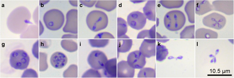
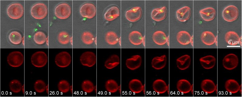
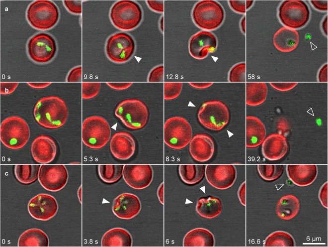
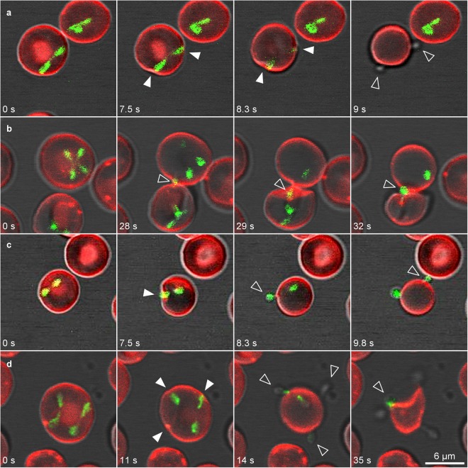
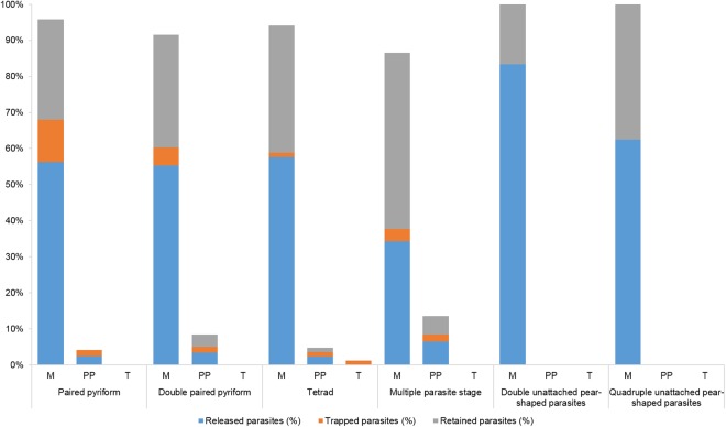
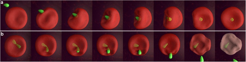
Based on confocal fluorescence and bright field video microscopy, we present detailed observations on the processes of invasion and egress of erythrocytes by the apicomplexan parasite Babesia divergens. Time-lapse images reveal numerous unexpected findings associated with the dynamics of B. divergens and its ability to manipulate the erythrocyte during both processes in its asexual cycle under in vitro conditions. Despite the speed at which these processes occur and the small size of the parasite, we capture infective merozoites moving vigorously and causing striking deformations in the erythrocyte's plasma membrane during an active invasion. We also observed intraerythrocytic dynamic stages as paired pyriforms, double paired pyriforms, tetrads, unattached pyriform sister cells and multiple parasite stages resulting in the release of large numbers of merozoites over a short period. Of considerable interest is that time-lapse images reveal a novel mechanism of egress used by B. divergens to exit the human erythrocyte. The release occurs when B. divergens parasites establish contacts with the plasma membrane of the erythrocyte from within, before exiting the cell. Visualization and analysis of the images enabled us to obtain useful information and broaden our knowledge of complex and crucial events involved with parasitisation of human erythrocytes by B. divergens.


Conflict of interest statement

The authors declare no competing interests.

Figures

Figure 1
Figure 1
B. divergens forms inside and outside erythrocytes were identified in in vitro cultures by Giemsa staining. (a) A free merozoite invading a human erythrocyte. Panels b–j show different stages within the human erythrocyte. (b) Single round trophozoite. (c) Paired pyriforms, a stage formed by two attached pear-shaped sister cells. (d) Double round trophozoites, (e) Double paired pyriforms. (f) Tetrad (g). Quadruple round trophozoites. (h) Multiple parasites. (i) Double unattached pyriforms. (j) Quadruple unattached pyriforms. (k) Intact paired pyriform outside the erythrocyte. (l) Intact tetrad outside the erythrocyte. Slides were examined with a Primo Star microscope (Zeiss, Germany) at 100X magnification.
Figure 2
Figure 2
The B. divergens free-merozoite recognizes and invades the human erythrocyte. The panels above show time-lapse image sequences of four B. divergens merozoites stained with MitoTracker (green fluorescent) that are exiting in 9 s from an infected human erythrocyte stained with PKH26 (red fluorescent). One of the merozoites identifies a new erythrocyte and establishes a tight irreversible connection with the erythrocyte plasma membrane causing a deep invagination and deformation to the erythrocyte at 48–75 s. The total internalization of the free merozoite occurs at ≈93 s. The parasite is surrounded by the red fluorescent erythrocyte membrane-derived parasitophorous vacuolar membrane after invasion. Simultaneously, the panels below show the PKH26 fluorescence around the infected and non-infected erythrocytes but also around the invasive parasite within the erythrocyte. Time-lapse imaging was captured every 1 s. The time-lapse between each frame is indicated in seconds.
Figure 3
Figure 3
B. divergens intraerythrocytic stages use similar strategies to exit from the infected erythrocyte. Time-lapse image sequences of the egress process of B. divergens parasites stained with MitoTracker (green fluorescent) from infected erythrocytes stained with PKH26 (red fluorescent). (a) A merozoite appears to emanate after the dramatic invagination (white arrowheads) of the erythrocyte plasma membrane provoked by the paired pyriform at 9.8–12.8 s. Seconds later, one free merozoite (arrowhead) is released from the erythrocyte while the other one remains within the host cell. (b) Egress process of a double paired pyriform. Both paired pyriforms produce successive dramatic depressions (white arrowheads) of the erythrocyte plasma membrane after 5.3 s and 8.3 s causing the discharge of one free merozoite (arrowhead) and the deformation of the erythrocyte. The other three merozoites are retained within or trapped outside the erythrocyte. (c) A tetrad causes an initial invagination in the erythrocyte plasma membrane (white arrowheads) at 3.8 sec. The depression and deformation of the erythrocyte become even clearer at 6 s. The tetrad can dissociate into four merozoites inside the cell. Only one free merozoite (arrowhead) is being released from the infected erythrocyte. Time-lapse imaging was captured every 0.753 s. The time-lapsed between each frame is indicated in seconds.
Figure 4
Figure 4
B. divergens merozoites exit the erythrocyte. Time-lapse image sequences of B. divergens parasites stained with MitoTracker (green fluorescent) and erythrocytes stained with PKH26 (red fluorescent). (a) A paired pyriform produces simultaneously depressions (white arrowheads) on two opposite sides of the erythrocyte plasma membrane at 7.5–8.3 s. Then, the two sister cells comprising the paired pyriform exert enough force in opposite directions to detach from each other and the detachment process results in two merozoites (arrowheads) in 8.3 s. Finally, both merozoites are simultaneously released from the opposite sides of the infected erythrocytes in 9 s. (b) An unattached merozoite from a tetrad touches the erythrocyte plasma membrane and is released (arrowhead) in 28 s following by two merozoites (arrowheads) that leave the cell from the same side in 29 s and 32 s, respectively. One merozoite remains within the erythrocyte. (c) Erythrocyte plasma membrane invagination (white arrowheads), causing by a paired pyriform at 7.5 s, facilitates the release of the two merozoites (arrowheads) from opposite sites. The first merozoite is released from the erythrocyte in 8.3 s and the second one in 9.8 s. (d) Two paired pyriforms (double paired pyriforms) simultaneously produces depressions on three different sides (white arrowheads) of the erythrocyte plasma membrane at 11 s. Then, four merozoites (arrowheads) are released from different sides at 14–35 s from the infected erythrocyte. Time-lapse imaging was captured every 0.753 or 1 s. The time-lapsed between each frame is indicated in seconds.
Figure 5
Figure 5
The egress efficiency of B. divergens was determined by the rate of parasite yield by the intraerythrocytic stages during the process. The graph shows the percentage of released, trapped and retained parasites within the infected erythrocytes: merozoite (M), paired pyriforms (PP), tetrads (T), n values were: n = 88 paired pyriforms, n = 71 double paired pyriforms, n = 23 tetrads, n = 76 multiple parasites stages, n = 3 double unattached pyriform parasites, n = 4 quadruple unattached pyriform parasites.
Figure 6
Figure 6
Serial diagram showing the most representative steps of the invasion and egress process of B. divergens. (a) The merozoite contact with the erythrocyte is vigorous and cause a deep invagination and waves of deformation on the erythrocyte membrane plasma during the entry. (b) Serial events represent a novel mechanism of egress used by B. divergens to exit the human erythrocyte. The intraerythrocytic parasite attaches to the erythrocyte to internalize the erythrocyte plasma membrane, a strategy that could be used by the parasites to exit the erythrocyte.

References

    1. Hildebrandt A, Gray JS, Hunfeld KP. Human babesiosis in Europe: what clinicians need to know. Infection. 2013;41:1057–1072. doi: 10.1007/s15010-013-0526-8. - DOI - PubMed
    1. Gray J, Zintl A, Hildebrandt A, Hunfeld KP, Weiss L. Zoonotic babesiosis: overview of the disease and novel aspects of pathogen identity. Ticks Tick Borne Dis. 2010;1:3–10. doi: 10.1016/j.ttbdis.2009.11.003. - DOI - PubMed
    1. Zintl A, Mulcahy G, Skerrett HE, Taylor SM, Gray JS. Babesia divergens, a bovine blood parasite of veterinary and zoonotic importance. Clin. Microbiol. Rev. 2003;16:622–636. doi: 10.1128/CMR.16.4.622-636.2003. - DOI - PMC - PubMed
    1. Hunfeld KP, Hildebrandt A, Gray JS. Babesiosis: recent insights into an ancient disease. Int. J. Parasitol. 2008;38:1219–1237. doi: 10.1016/j.ijpara.2008.03.001. - DOI - PubMed
    1. Sun Y, Moreau E, Chauvin A, Malandrin L. The invasion process of bovine erythrocyte by Babesia divergens: knowledge from an in vitro assay. Vet. Res. 2011;42:62. doi: 10.1186/1297-9716-42-62. - DOI - PMC - PubMed

Publication types

LinkOut - more resources