. 2018 Sep 7:13:5231-5248.
doi: 10.2147/IJN.S167142. eCollection 2018.

Mesenchymal stem cells loaded with paclitaxel-poly(lactic- co-glycolic acid) nanoparticles for glioma-targeting therapy

Affiliations

Mesenchymal stem cells loaded with paclitaxel-poly(lactic- co-glycolic acid) nanoparticles for glioma-targeting therapy

Xiaoling Wang et al. Int J Nanomedicine. .

Abstract

Background: Mesenchymal stem cells (MSCs) possess inherent tropism towards tumor cells, and so have attracted increased attention as targeted-therapy vehicles for glioma treatment.

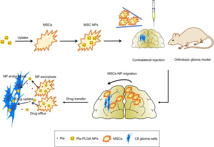
Purpose: The objective of this study was to demonstrate the injection of MSCs loaded with paclitaxel (Ptx)-encapsulated poly(d,l-lactide-co-glycolide) (PLGA) nanoparticles (NPs) for orthotopic glioma therapy in rats.

Methods: Ptx-PLGA NP-loaded MSC was obtained by incubating MSCs with Ptx-PLGA NPs. The drug transfer and cytotoxicity of Ptx-PLGA NP-loaded MSC against tumor cells were investigated in the transwell system. Biodistribution and antitumor activity was evaluated in the orthotopic glioma rats after contralateral injection.

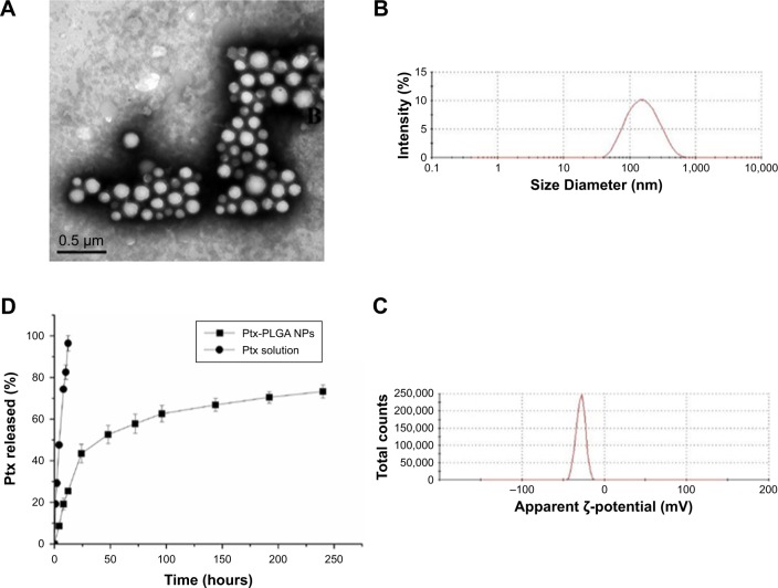
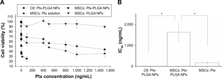
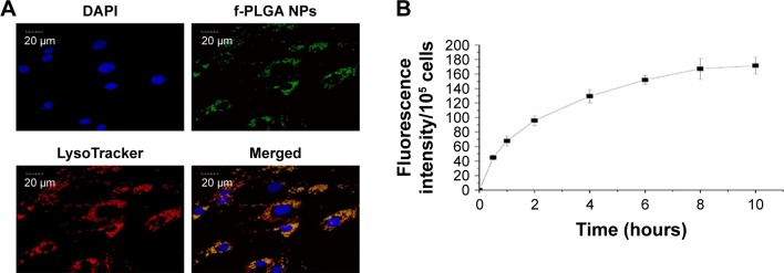
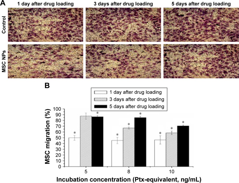
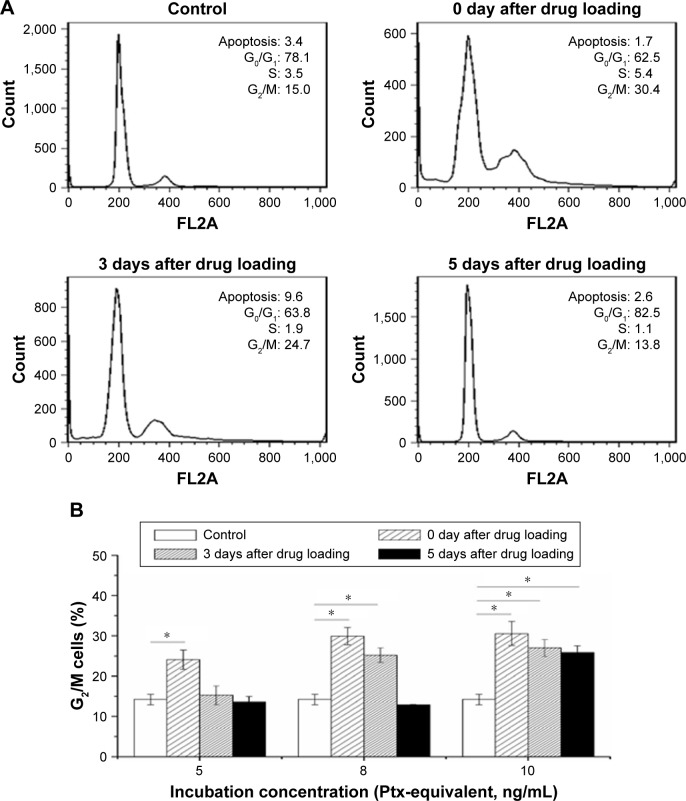
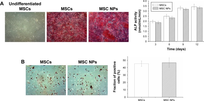
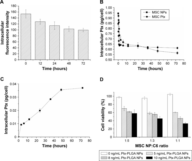
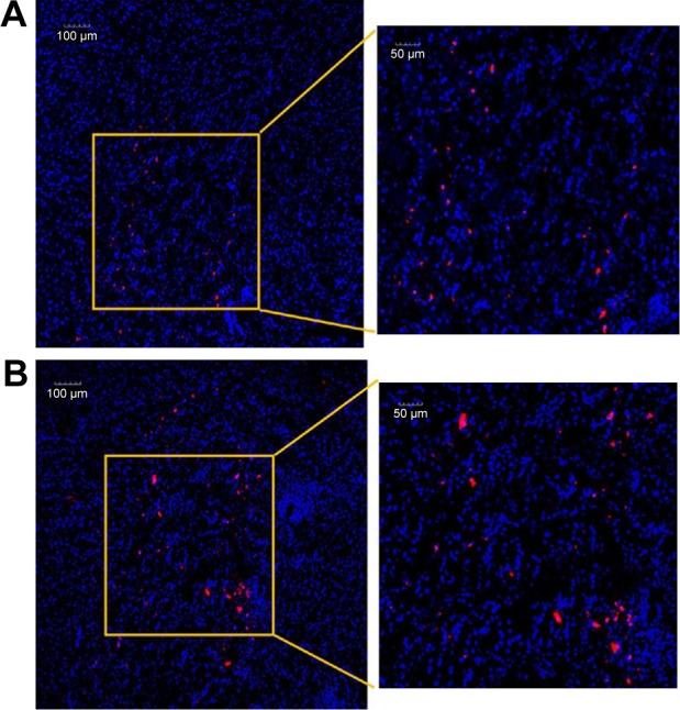
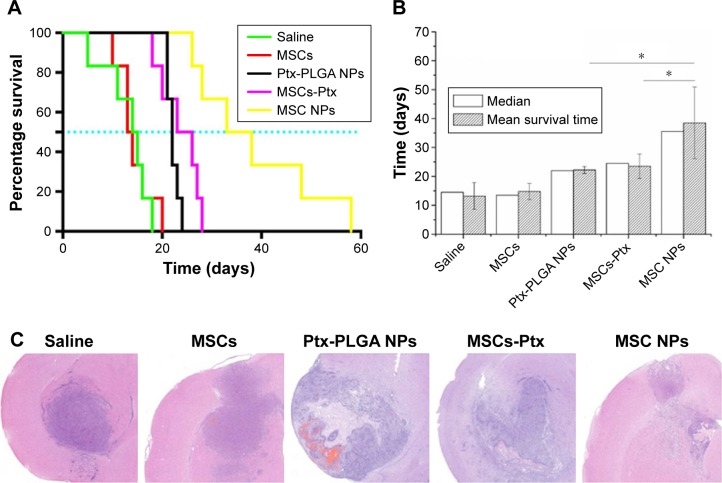
Results: The optimal dose of MSC-loaded Ptx-PLGA NPs (1 pg/cell Ptx) had little effect on MSC-migration capacity, cell cycle, or multilineage-differentiation potential. Compared with Ptx-primed MSCs, Ptx-PLGA NP-primed MSCs had enhanced sustained Ptx release in the form of free Ptx and Ptx NPs. Ptx transfer from MSCs to glioma cells could induce tumor cell death in vitro. As for distribution in vivo, NP-loaded fluorescent MSCs were tracked throughout the tumor mass for 2 days after therapeutic injection. Survival was significantly longer after contralateral implantation of Ptx-PLGA NP-loaded MSCs than those injected with Ptx-primed MSCs or Ptx-PLGA NPs alone.

Conclusion: Based on timing and sufficient Ptx transfer from the MSCs to the tumor cells, Ptx-PLGA NP-loaded MSC is effective for glioma treatment. Incorporation of chemotherapeutic drug-loaded NPs into MSCs is a promising strategy for tumor-targeted therapy.

Keywords: BMSCs; C6 cells; contralateral injection; drug targeting; orthotopic glioma.


Conflict of interest statement

Disclosure The authors report no conflicts of interest in this work.

Figures

Figure 1
Figure 1
Glioma-targeting delivery of MSCs loaded with Ptx-PLGA NPs. Notes: MSCs-NPs were prepared by incubation MSCs with Ptx-PLGA NPs. Then MSCs-NPs migrate into glioma and sustained release of Ptx after contralateral injection. Abbreviations: MSCs, mesenchymal stem cells; Ptx, paclitaxel; PLGA, poly(d,l-lactide-co-glycolide); NPs, nanoparticles.
Figure 2
Figure 2
Characteristics of Ptx-PLGANPs. Notes: (A) Transmission electron microscopy of Ptx-PLGANPs; (B) size distribution of Ptx-PLGANPs; (C) ζ-potential of Ptx-PLGANPs; (D) in vitro release of Ptx from Ptx-PLGANPs. Abbreviations: Ptx, paclitaxel; PLGA, poly(d,l-lactide-co-glycolide); NPs, nanoparticles.
Figure 3
Figure 3
Cytotoxicity of Ptx-PLGA NPs against MSCs and C6 glioma cells. Notes: (A) Cell viability determined by MTT assay after incubation with Ptx-PLGA NPs, Ptx solution, and blank PLGA NPs for 72 hours. (B) IC50 values of Ptx-PLGA NPs and Ptx solution in C6 cells and MSCs *(P<0.05). Abbreviations: Ptx, paclitaxel; PLGA, poly(d,l-lactide-co-glycolide); NPs, nanoparticles; MSCs, mesenchymal stem cells.
Figure 4
Figure 4
Intracellular distribution and internalization kinetics of f-PLGA NPs in MSCs. Notes: (A) Confocal laser-scanning microscopy of MSCs treated with f-PLGA NPs (5 µg/mL). Lysosomes and nuclei were stained with LysoTracker Red and DAPI, respectively. (B) Uptake kinetics of f-PLGA NPs into MSCs. Abbreviations: f-PLGA NPs, fluorescein–poly(d,l-lactide-co-glycolide) nanoparticles; MSCs, mesenchymal stem cells.
Figure 5
Figure 5
Restoration of migratory activity of MSC NPs in vitro. Notes: (A) Representative photographs of migrated MSCs through the membrane pores. MSCs were transferred to the upper chamber of the transwell system after exposure to 8 ng/mL Ptx-PLGA NPs for 8 hours. At 1, 3, and 5 days later, migrated MSCs were stained by crystal violet (400×). (B) Percentage of MSC NPs migrating. MSCs were treated with 5, 8, and 10 ng/mL Ptx-PLGA NPs for 8 hours, then cells were washed and seeded onto the transwell system. Numbers of migrating MSCs at 1, 3, and 5 days after seeding were counted. Cells incubated with blank NPs were considered the control *(P<0.05 compared with the number of migrated cells in control group at different time points after drug loading). Abbreviations: MSC NPs, mesenchymal stem cells loaded with Ptx-PLGA NPs; Ptx, paclitaxel; PLGA, poly(d,l-lactide-co-glycolide); NPs, nanoparticles.
Figure 6
Figure 6
Cell-cycle progression restoration of MSC NPs from Ptx-induced cell-cycle arrest. Notes: (A) Cell-cycle analysis of MSCs recovered from exposure to 8 ng/mL Ptx-PLGA NPs; (B) percentage of MSCs arrested in G2/M phase 0, 3, and 5 days after incubation with 5, 8, and 10 ng/mL Ptx-PLGA NPs *(P<0.05). Abbreviations: MSC, mesenchymal stem cell; NPs, nanoparticles; Ptx, paclitaxel; PLGA, poly(d,l-lactide-co-glycolide).
Figure 7
Figure 7
Osteogenic and adipogenic differentiation of MSCs treated with 8 ng/mL Ptx-PLGA NPs for 8 hours. Notes: (A) Osteogenic differentiation of MSC NPs. Calcium-nodule deposits were stained with alizarin red. Undifferentiated MSCs were set as the negative control. Quantification of ALP was performed in the early stage of osteogenic differentiation. (B) Adipogenic differentiation of MSC NPs. Neutral lipid vacuoles were stained with oil red O (400×). The fraction of positive cells was compared with that of untreated MSCs. Abbreviations: MSCs, mesenchymal stem cells; Ptx, paclitaxel; PLGA, poly(d,l-lactide-co-glycolide); NPs, nanoparticles.
Figure 8
Figure 8
Ptx transferred from MSC NPs to C6 cells. Notes: (A) Exocytosis of f-PLGA NPs from MSCs. (B) Kinetics of Ptx released from MSC NPs. (C) Uptake kinetics of Ptx by C6 cells in the transwell system. MSC NPs were seeded in the upper chamber. (D) Antitumor effects of MSC NPs against C6 cells in the transwell system. Abbreviations: Ptx, paclitaxel; f-PLGA NPs, fluorescein-poly(d,l-lactide-co-glycolide) nanoparticles; MSC NPs, mesenchymal stem cells loaded with Ptx-PLGA NPs.
Figure 9
Figure 9
Intracerebral distribution of MSC NPs. Notes: (A) Confocal laser-scanning microscopy of glioma. All the area was within the glioma. (B) Brain tissue at the therapeutic injection site 2 days after contralateral injection of MSC NPs. MSCs were incubated with 8 ng/mL Ptx-PLGA NPs for 8 hours, and labeled with a red fluorescent probe – CM-Dil. Nuclei were stained with DAPI before observation. Abbreviations: Ptx-PLGA NPs, paclitaxel poly(d,l-lactide-co-glycolide) nanoparticles; MSC, mesenchymal stem cell.
Figure 10
Figure 10
In vivo antitumor efficacy of MSC NPs. Notes: (A) Kaplan–Meier survival curve; (B) median survival time *(P<0.05); (C) H&E sections of tumor tissues from C6 glioma-bearing rats. Ptx-PLGA NPs, MSCs-Ptx, and MSCs-NPs with a dose of 1 µg Ptx-equivalent/kg, as well as saline and MSC controls, were injected contralaterally 1 week after tumor implantation. On the seventh day after therapeutic injection, tumor masses were stained with H&E for histopathology evaluation. Abbreviations: Ptx-PLGA NPs, paclitaxel poly(d,l-lactide-co-glycolide) nanoparticles; MSC, mesenchymal stem cell.

Similar articles

Cited by

References

    1. Chang L, Su J, Jia X, Ren H. Treating malignant glioma in Chinese patients: update on temozolomide. Onco Targets Ther. 2014;7:235–244. - PMC - PubMed
    1. Zhang TY, Huang B, Yuan ZY, et al. Gene recombinant bone marrow mesenchymal stem cells as a tumor-targeted suicide gene delivery vehicle in pulmonary metastasis therapy using non-viral transfection. Nanomedicine. 2014;10(1):257–267. - PubMed
    1. Zhang TY, Huang B, Wu HB, et al. Synergistic effects of co-administration of suicide gene expressing mesenchymal stem cells and prodrug-encapsulated liposome on aggressive lung melanoma metastases in mice. J Control Release. 2015;209:260–271. - PubMed
    1. Choi SA, Lee YE, Kwak PA, et al. Clinically applicable human adipose tissue-derived mesenchymal stem cells delivering therapeutic genes to brainstem gliomas. Cancer Gene Ther. 2015;22(6):302–311. - PubMed
    1. Bexell D, Svensson A, Bengzon J. Stem cell-based therapy for malignant glioma. Cancer Treat Rev. 2013;39(4):358–365. - PubMed

MeSH terms