

Mycoplasma hominis empyema following caesarean section
- PMID: 30237888
- PMCID: PMC6138538
- DOI: 10.1002/rcr2.367
Mycoplasma hominis empyema following caesarean section
Abstract
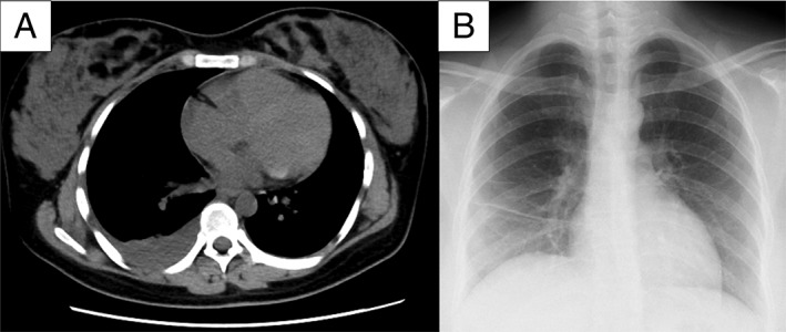
Mycoplasma hominis as a cause of empyema is rare. We report a case of empyema caused by M. hominis following a caesarean section. A 28-year-old woman at 39 weeks and one day of pregnancy was admitted to our hospital and underwent an emergency caesarean section because of premature rupture of membranes. On postoperative day 2, she developed a fever, and flomoxef was administered. A pleural effusion developed on the right side. A diagnosis of empyema was made, and sulbactam/ampicillin was administered. However, the patient's clinical condition did not improve. Numerous small pinpoint colonies, which did not yield visible bacteria on a Gram stain, were observed on a plate of pleural fluid culture, and M. hominis empyema was suspected. Based on this result, antibiotic therapy was switched to clindamycin, and the patient's clinical condition improved rapidly. M. hominis was detected in the pleural fluid by polymerase chain reaction (PCR) assay. M. hominis should be considered a causative pathogen for empyema following a caesarean section.
Keywords: Bloodstream infection; Mycoplasma hominis; caesarean section; empyema; pregnancy.
Figures

Similar articles
-
[Mycoplasma hominis empyema following pleuropneumonia in late pregnancy].Schweiz Med Wochenschr. 1993 Nov 27;123(47):2244-6. Schweiz Med Wochenschr. 1993. PMID: 8272796 German.
-
Pelvic abscess due to Mycoplasma hominis following caesarean section.JMM Case Rep. 2016 Aug 30;3(4):e005059. doi: 10.1099/jmmcr.0.005059. eCollection 2016 Aug. JMM Case Rep. 2016. PMID: 28348780 Free PMC article.
-
Mycoplasma hominis Intracranial Abscess Diagnosed by Characteristic Colonies Obtained Through Extended Culture: Case Report and Literature Review.Cureus. 2025 Mar 3;17(3):e79981. doi: 10.7759/cureus.79981. eCollection 2025 Mar. Cureus. 2025. PMID: 40034420 Free PMC article.
-
Postoperative Mycoplasma hominis infections after neurosurgical intervention.J Neurosurg Pediatr. 2014 Aug;14(2):212-8. doi: 10.3171/2014.4.PEDS13547. Epub 2014 May 23. J Neurosurg Pediatr. 2014. PMID: 24856879 Review.
-
Meningitis in a Chinese adult patient caused by Mycoplasma hominis: a rare infection and literature review.BMC Infect Dis. 2016 Oct 12;16(1):557. doi: 10.1186/s12879-016-1885-4. BMC Infect Dis. 2016. PMID: 27729031 Free PMC article. Review.
Cited by
-
Bloodstream Infection Combined with Thoracic Infection Caused by Mycoplasma hominis: A Case Report and Review of the Literature.Infect Drug Resist. 2024 Dec 24;17:5795-5801. doi: 10.2147/IDR.S478555. eCollection 2024. Infect Drug Resist. 2024. PMID: 39734739 Free PMC article.
-
Application of next-generation sequencing on diagnosis of bloodstream infection caused by Mycoplasma hominis in a patient with ANCA-associated vasculitis.Ann Clin Microbiol Antimicrob. 2023 Apr 21;22(1):28. doi: 10.1186/s12941-023-00580-4. Ann Clin Microbiol Antimicrob. 2023. PMID: 37085831 Free PMC article.
References
-
- Lyon GM, Alspaugh JA, Meredith FT, et al. 1997. Mycoplasma hominis pneumonia complicating bilateral lung transplantation: case report and review of the literature. Chest 112:1428–1432. - PubMed
-
- Taylor‐Robinson D. 1995. Ureaplasma urealyticum and mycoplasma hominis Pp. 1713–1717 in Mandell C. L., Bennett J. E., Dolin R., eds. Principles and practice of infectious diseases. New York, NY, Churchill Livingstone.
-
- Fabbri J, Tamm M, Frei R, et al. 1993. Mycoplasma hominis empyema following pleuropneumonia in late pregnancy. Schweiz. Med. Wochenschr. 123:2244–2246. - PubMed
Publication types
LinkOut - more resources
Full Text Sources
Other Literature Sources
