. 2016 Mar 23;6(1):e12.
doi: 10.2106/JBJS.ST.15.00065.

Isolated Subtalar Arthrodesis

Affiliations

Isolated Subtalar Arthrodesis

Paulo N Ferrao et al. JBJS Essent Surg Tech. .

Abstract

Introduction: An isolated subtalar arthrodesis through a sinus tarsi approach with cannulated screw fixation is safe, reproducible, and effective for specific hindfoot pathology in adults.

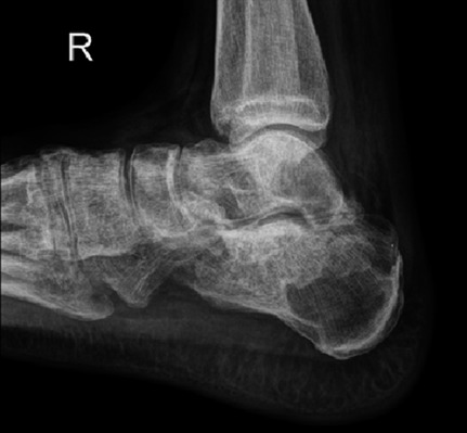
Step 1 preoperative planning: Perform a comprehensive clinical and radiographic assessment.

Step 2 patient positioning: Position the patient supine on the operating table with a bump under the ipsilateral hip, to internally rotate the involved leg.

Step 3 incision: Mark a longitudinal incision from just inferior to the tip of the lateral malleolus and extending toward the base of the 4th metatarsal.

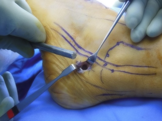
Step 4 approach: Expose the subtalar joint by reflecting the extensor digitorum brevis and protecting the peroneal tendons.

Step 5 joint preparation: Meticulously prepare the subtalar joint using osteotomes and curets down to healthy bleeding subchondral bone.

Step 6 reduction and fixation: Reduce the subtalar joint into 5° of valgus and fix it with cannulated screws.

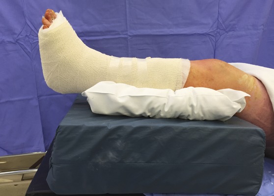
Step 7 wound closure: Meticulously close the wound in layers and place the lower leg in a well-padded plaster back slab with the ankle in a neutral position.

Step 8 postoperative care: The operatively treated leg is kept immobilized and non-weight-bearing for the first 6 weeks.

Results: Numerous retrospective articles have reported high fusion rates and good functional outcomes after isolated subtalar arthrodesis, for all indications.


Figures

Fig. 1
Fig. 1
Hindfoot valgus malalignment of the left foot as seen from behind the patient.
Fig. 2
Fig. 2
Weight-bearing oblique (Fig. 2-A), weight-bearing anteroposterior (Fig. 2-B), and weight-bearing lateral (Fig. 2-C) radiographs of a patient with isolated subtalar arthritis.
Fig. 3
Fig. 3
Saltzman view for measuring hindfoot alignment relative to the tibia as described by Donovan and Rosenberg.
Fig. 4
Fig. 4
The patient is positioned supine with the feet at the edge of the table and a bump under the ipsilateral hip.
Fig. 5
Fig. 5
Two rolled towels are placed under the heel to lift the foot off the table, making surgical access easier.
Fig. 6
Fig. 6
The surgical incision is drawn from below the tip of the lateral malleolus to the base of the 4th metatarsal. The superficial peroneal and sural nerves are drawn as dotted lines.
Fig. 7
Fig. 7
The origin of the extensor digitorum brevis is identified and released. The muscle belly is dissected out distally and sutured to the distal skin.
Fig. 8
Fig. 8
A Hohmann retractor is placed deep to the calcaneofibular ligament around the posterior aspect of the subtalar joint, improving exposure and protecting the peroneal tendons.
Fig. 9
Fig. 9
A lamina spreader is used to open the joint, making access for debridement easier.
Fig. 10
Fig. 10
The flexor hallucis longus tendon is identified in the posteromedial aspect of the subtalar joint. It is important not to damage the tendon with osteotomes during debridement.
Fig. 11-A
Fig. 11-A
Autogenous bone graft being taken from the calcaneus.
Fig. 11-B
Fig. 11-B
Autogenous bone graft being taken from the distal part of the tibia. The incision is made 2 cm proximal to the tip of the medial malleolus.
Fig. 11-C
Fig. 11-C
Autogenous bone graft from the distal part of the tibia.
Fig. 12
Fig. 12
The incision marked on the posterior aspect of the heel, off the weight-bearing surface of the foot, for insertion of the screws.
Fig. 13
Fig. 13
Fully threaded conical headless compression screws.
Fig. 14
Fig. 14
The guidewires are inserted through the heel into the talus while the joint is reduced.
Fig. 15-A
Fig. 15-A
Posttraumatic subtalar arthritis with loss of calcaneal height and decreased talar declination.
Fig. 15-B
Fig. 15-B
Subtalar bone-block distraction arthrodesis with restored calcaneal height and talar declination.
Fig. 16-A
Fig. 16-A
Lateral fluoroscopic view to confirm that the guidewires were inserted in a divergent pattern and crossing the subtalar joint.
Fig. 16-B
Fig. 16-B
Anteroposterior fluoroscopic view of the ankle confirming that the guidewires are within the talus.
Fig. 17
Fig. 17
Intraoperative fluoroscopy confirming correct screw length and placement.
Fig. 18
Fig. 18
The extensor digitorum brevis muscle is sutured back to its origin.
Fig. 19-A
Fig. 19-A
A back slab is applied using 2 plaster splints over a bulky dressing.
Fig. 19-B
Fig. 19-B
The operatively treated leg immobilized in the below-the-knee plaster slab, with the ankle in a neutral position, and elevated.
Fig. 20
Fig. 20
Radiograph made at 16 weeks confirming subtalar fusion.

References

    1. Greisberg J, Sangeorzan B. Hindfoot arthrodesis. J Am Acad Orthop Surg. 2007. January;15(1):65-71. - PubMed
    1. Mann RA. Arthrodesis of the foot and ankle. In: Coughlin MJ, Mann RA, Saltzman CL, editors. Surgery of the foot and ankle. 8th ed Philadelphia: Elsevier Health; 2007. p 1087-123.
    1. Astion DJ, Deland JT, Otis JC, Kenneally S. Motion of the hindfoot after simulated arthrodesis. J Bone Joint Surg Am. 1997. February;79(2):241-6. - PubMed
    1. Roster B, Kreulen C, Giza E. Subtalar joint arthrodesis: open and arthroscopic indications and surgical techniques. Foot Ankle Clin. 2015. June;20(2):319-34. Epub 2015 Apr 11. - PubMed
    1. Hentges MJ, Gesheff MG, Lamm BM. Realignment subtalar joint arthrodesis. J Foot Ankle Surg. 2016. Jan-Feb;55(1):16-21. Epub 2015 May 28. - PubMed

LinkOut - more resources