

Case Reports
doi: 10.1016/j.jdcr.2018.05.016.
eCollection 2018 Sep.
55-year-old man with ulcers in inguinal fold and intergluteal cleft found to have systemic Langerhans cell histiocytosis
Affiliations
- PMID: 30238052
- PMCID: PMC6143693
- DOI: 10.1016/j.jdcr.2018.05.016
Item in Clipboard
Case Reports
55-year-old man with ulcers in inguinal fold and intergluteal cleft found to have systemic Langerhans cell histiocytosis
JAAD Case Rep.
.
No abstract available
Keywords: BRAF V600 mutations; Birbeck granules; LCH, Langerhans cell histiocytosis; Langerhans cell histiocytosis; ulcers.
Figures
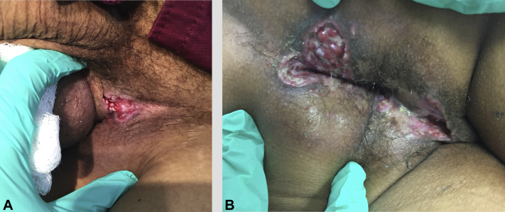
A and B, Well-demarcated inguinal and intergluteal ulcers found during physicial examination were nontender on palpation.
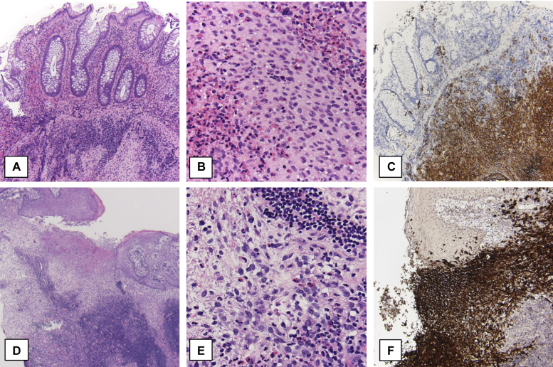
Colon and skin histopathology of patient with Langerhans cell histiocytosis. A, Polypoid colonic lesion shows a dense proliferation of histiocytes in the submucosa with eosinophilic abscesses in addition to neutrophilic and lymphocytic infiltrates. B, Oval histiocytes approximately 15 μm characterized by folded, indented or lobulated nuclei with fine chromatin, inconspicuous nucleoli, and thin nuclear membranes. The cytoplasms are moderately abundant and slightly eosinophilic. C, Langerhans cell histiocytosis involvement at colon. D, Skin biopsy reveals similar infiltrate to that of the colon specimen. E, Histiocytes with large, pale, folded or lobulated (often reniform), vesicular nuclei and abundant, slightly eosinophilic or amphophilic cytoplasms. F, Histiocytic infiltrate. (A, B, D, and E; Hematoxylin-eosin stain; C and F; CD1a stain; original magnification: A, C, D, and F, X2; B and E, X20.)
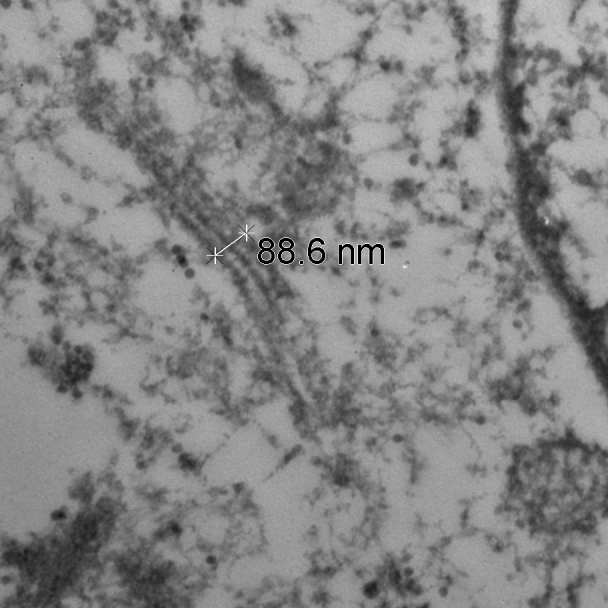
Electron microscopy image of cytoplasm of Langerhans cell shows characteristic Birbeck granules.
Similar articles
-
BRAF V600 mutations in Langerhans cell histiocytosis with a simple and unique assay.Diagn Pathol. 2016 Apr 19;11:39. doi: 10.1186/s13000-016-0489-z. Diagn Pathol. 2016. PMID: 27094161 Free PMC article.
-
Pediatric Langerhans cell histiocytosis: the impact of mutational profile on clinical progression and late sequelae.Ann Hematol. 2019 Jul;98(7):1617-1626. doi: 10.1007/s00277-019-03678-y. Epub 2019 Mar 28. Ann Hematol. 2019. PMID: 30923995
-
Recurrent NRAS mutations in pulmonary Langerhans cell histiocytosis.Eur Respir J. 2016 Jun;47(6):1785-96. doi: 10.1183/13993003.01677-2015. Epub 2016 Apr 13. Eur Respir J. 2016. PMID: 27076591
-
BRAF gene mutations in synchronous papillary thyroid carcinoma and Langerhans cell histiocytosis co-existing in the thyroid gland: a case report and literature review.BMC Cancer. 2019 Feb 22;19(1):170. doi: 10.1186/s12885-019-5372-3. BMC Cancer. 2019. PMID: 30795755 Free PMC article. Review.
-
[Status of Langerhans cell histiocytosis in children and adults].Rinsho Ketsueki. 2019;60(9):1308-1316. doi: 10.11406/rinketsu.60.1308. Rinsho Ketsueki. 2019. PMID: 31597857 Review. Japanese.
Cited by
-
Langerhans cell histiocytosis in an adult female with atypical swellings.Indian J Dermatol Venereol Leprol. 2021 Mar-Apr;87(2):254-259. doi: 10.25259/IJDVL_651_19. Indian J Dermatol Venereol Leprol. 2021. PMID: 33769735 No abstract available.
-
Skin-limited Langerhans cell histiocytosis in an adult presenting as isolated, eroded, "kissing" intergluteal plaques.JAAD Case Rep. 2023 Oct 1;42:16-19. doi: 10.1016/j.jdcr.2023.09.019. eCollection 2023 Dec. JAAD Case Rep. 2023. PMID: 37965193 Free PMC article. No abstract available.
References
-
- Baumgartner I., von Hochstetter A., Baumert B., Luetolf U., Follath F. Langerhans'-cell histiocytosis in adults. Med Pediatr Oncol. 1997;28:9–14. - PubMed
-
- Osband M.E. Histiocytosis X. Langerhans' cell histiocytosis. Hematol Oncol Clin North Am. 1987;1:737–751. - PubMed
-
- Chu T., D'Angio G.J., Favara B.E., Ladisch S., Nesbit M., Pritchard J. Histiocytosis syndromes in children. Lancet. 1987;2:41–42. - PubMed
-
- Enriquez P., Dahlin D.C., Hayles A.B., Henderson E.D. Histiocytosis X: a clinical study. Mayo Clin Proc. 1967;42:88–99. - PubMed
-
- Malpas J.S. Langerhans cell histiocytosis in adults. Hematol Oncol Clin North Am. 1998;12:259–268. - PubMed
Publication types
Grants and funding
LinkOut - more resources
Full Text Sources
Other Literature Sources
Research Materials