

Cerebrospinal fluid volumetric net flow rate and direction in idiopathic normal pressure hydrocephalus
- PMID: 30238917
- PMCID: PMC6154456
- DOI: 10.1016/j.nicl.2018.09.006
Cerebrospinal fluid volumetric net flow rate and direction in idiopathic normal pressure hydrocephalus
Abstract
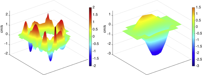
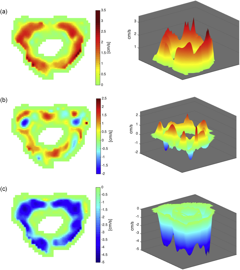
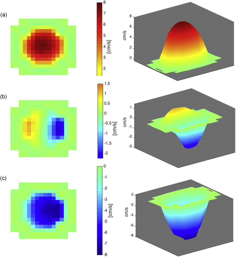
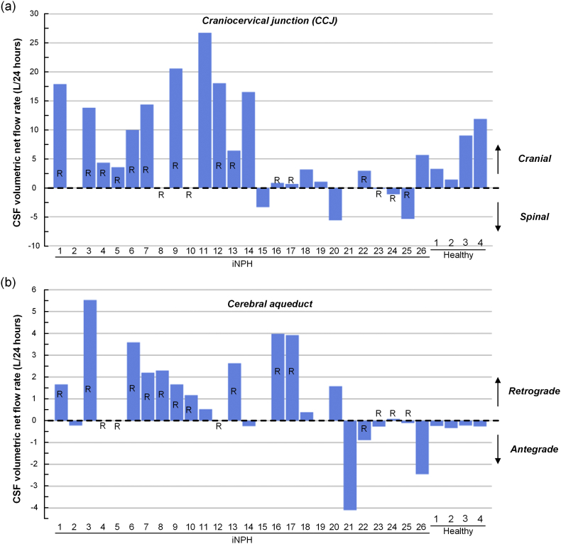
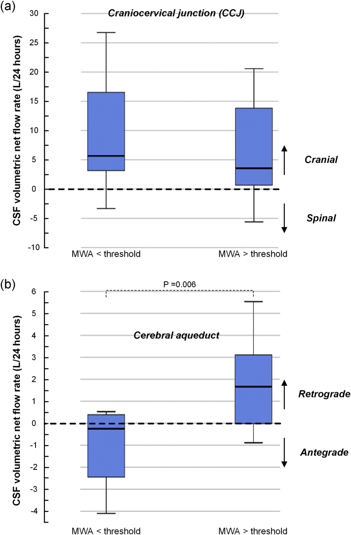
The aim of the present study was to examine cerebrospinal fluid (CSF) volumetric net flow rate and direction at the cranio-cervical junction (CCJ) and cerebral aqueduct in individuals with idiopathic normal pressure hydrocephalus (iNPH) using cardiac-gated phase-contrast magnetic resonance imaging (PC-MRI). An in-depth, pixel-by-pixel analysis of regions of interest from the CCJ and cerebral aqueduct, respectively, was done in 26 iNPH individuals, and in 4 healthy subjects for validation purposes. Results from patients were compared with over-night measurements of static and pulsatile intracranial pressure (ICP). In iNPH, CSF net flow at CCJ was cranially directed in 17/22 as well as in 4/4 healthy subjects. Estimated daily CSF volumetric net flow rate at CCJ was 6.9 ± 9.9 L/24 h in iNPH patients and 4.5 ± 5.0 L/24 h in healthy individuals. Within the cerebral aqueduct, the CSF net flow was antegrade in 7/21 iNPH patients and in 4/4 healthy subjects, while it was retrograde (i.e. towards ventricles) in 14/21 iNPH patients. Estimated daily CSF volumetric net flow rate in cerebral aqueduct was 1.1 ± 2.2 L/24 h in iNPH while 295 ± 53 mL/24 h in healthy individuals. Magnitude of cranially directed CSF net flow in cerebral aqueduct was highest in iNPH individuals with signs of impaired intracranial compliance. The study results indicate CSF flow volumes and direction that are profoundly different from previously assumed. We hypothesize that spinal CSF formation may serve to buffer increased demand for CSF flow through the glymphatic system during sleep and during deep inspiration to compensate for venous outflow.
Keywords: Cerebral aqueduct; Cerebrospinal fluid; Cranio-cervical junction; Intracranial pressure; Phase-contrast MRI.
Copyright © 2018 The Author(s). Published by Elsevier Inc. All rights reserved.
Figures

References
-
- Baledent O., Gondry-Jouet C., Meyer M.E., De Marco G., Le Gars D., Henry-Feugeas M.C., Idy-Peretti I. Relationship between cerebrospinal fluid and blood dynamics in healthy volunteers and patients with communicating hydrocephalus. Investig. Radiol. 2004;39:45–55. - PubMed
-
- Bateman G.A., Brown K.M. The measurement of CSF flow through the aqueduct in normal and hydrocephalic children: from where does it come, to where does it go? Childs Nerv. Syst. 2012;28:55–63. - PubMed
-
- Bedussi B., van der Wel N.N., de Vos J., van Veen H., Siebes M., Vanbavel E., Bakker E.N. Paravascular channels, cisterns, and the subarachnoid space in the rat brain: a single compartment with preferential pathways. J. Cereb. Blood Flow Metab. 2017 Apr;37(4):1374–1385. (Epub 2016 Jan 1) - PMC - PubMed
-
- Bering E.A., Jr. Water exchange of central nervous system and cerebrospinal fluid. J. Neurosurg. 1952;9:275–287. - PubMed
-
- Bradley W.G.,.Jr., Scalzo D., Queralt J., Nitz W.N., Atkinson D.J., Wong P. Normal-pressure hydrocephalus: evaluation with cerebrospinal fluid flow measurements at MR imaging. Radiology. 1996;198:523–529. - PubMed
Publication types
MeSH terms
LinkOut - more resources
Full Text Sources
Other Literature Sources
