

Typical asymmetry in the hemispheric activation during an fMRI verbal comprehension paradigm is related to better performance in verbal and non-verbal tasks in patients with epilepsy
- PMID: 30238918
- PMCID: PMC6154460
- DOI: 10.1016/j.nicl.2018.09.010
Typical asymmetry in the hemispheric activation during an fMRI verbal comprehension paradigm is related to better performance in verbal and non-verbal tasks in patients with epilepsy
Erratum in
-
Erratum to typical asymmetry in the hemispheric activation during an fMRI verbal comprehension paradigm is related to better performance in verbal and non-verbal tasks in patients with epilepsy. NeuroImage: Clinical 20 (2018) 742-752.Neuroimage Clin. 2019;21:101700. doi: 10.1016/j.nicl.2019.101700. Epub 2019 Feb 4. Neuroimage Clin. 2019. PMID: 30733109 Free PMC article. No abstract available.
Abstract
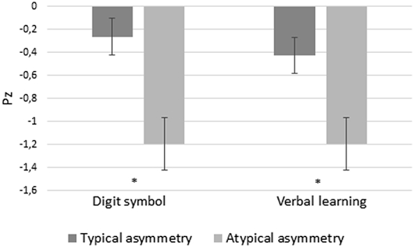
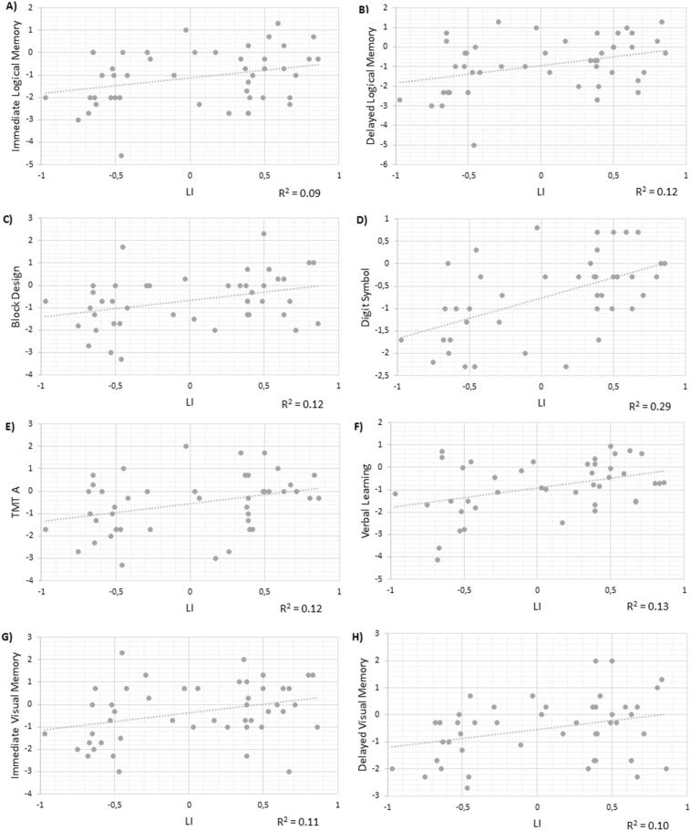
Chronic exposure to seizures in patients with left hemisphere (LH) epileptic focus could favor higher activation in the contralateral hemisphere during language processing, but the cognitive effects of this remain unclear. This study assesses the relationship between asymmetry in hemispheric activation during language fMRI and performance in verbal and non-verbal tasks. Whereas prior studies primarily used fMRI paradigms that favor frontal lobe activation and less prominent activation of the medial or superior temporal lobes, we used a verbal comprehension paradigm previously demonstrated to activate reliably receptive language areas. Forty-seven patients with drug-resistant epilepsy candidates for surgery underwent a multidisciplinary assessment, including a comprehensive neuropsychological evaluation and an fMRI verbal comprehension paradigm. Patients were distributed in two groups depending on laterality indexes (LI): typical hemispheric asymmetry (unilateral left activation preponderance; n = 23) and atypical hemispheric asymmetry (bilateral or unilateral right preponderance; n = 24). Right-handedness and right hemisphere (RH) focus were significant predictors of typical asymmetry. Patients with typical activation pattern presented better performance intelligence quotient and verbal learning than patients with atypical hemispheric asymmetry (for all, p < 0.014). Patients with LH focus had more frequently atypical hemispheric asymmetry than patients with RH focus (p = 0.05). Specifically, they showed lower LI and this was related to worse performance in verbal and non-verbal tasks. In conclusion, an increased activation of homologous RH areas for verbal comprehension processing could imply a competition of cognitive resources in the performance of the same task, disrupting cognitive performance.
Keywords: Cognitive performance; Epilepsy; Language; Typical asymmetry; fMRI.
Copyright © 2018 The Authors. Published by Elsevier Inc. All rights reserved.
Figures


Similar articles
-
Does education play a role in language reorganization after surgery in drug refractory temporal lobe epilepsy: An fMRI based study?Epilepsy Res. 2017 Oct;136:88-96. doi: 10.1016/j.eplepsyres.2017.07.017. Epub 2017 Jul 29. Epilepsy Res. 2017. PMID: 28802988
-
Seizure focus affects regional language networks assessed by fMRI.Neurology. 2005 Nov 22;65(10):1604-11. doi: 10.1212/01.wnl.0000184502.06647.28. Neurology. 2005. PMID: 16301489
-
Strengthening of laterality of verbal and visuospatial functions during childhood and adolescence.Hum Brain Mapp. 2009 Feb;30(2):473-83. doi: 10.1002/hbm.20523. Hum Brain Mapp. 2009. PMID: 18219619 Free PMC article.
-
Laterality of Brain Activation for Risk Factors of Addiction.Curr Drug Abuse Rev. 2016;9(1):1-18. doi: 10.2174/1874473709666151217121309. Curr Drug Abuse Rev. 2016. PMID: 26674074 Free PMC article. Review.
-
Gender and Hemispheric Asymmetries in Acquired Sociopathy.Front Psychol. 2019 Mar 19;10:346. doi: 10.3389/fpsyg.2019.00346. eCollection 2019. Front Psychol. 2019. PMID: 30941065 Free PMC article.
Cited by
-
Clinical practice of language fMRI in epilepsy centers: a European survey and conclusions by the ESNR Epilepsy Working Group.Neuroradiology. 2020 May;62(5):549-562. doi: 10.1007/s00234-020-02397-w. Epub 2020 Mar 13. Neuroradiology. 2020. PMID: 32170372 Free PMC article.
References
-
- Adcock J.E., Wise R.G., Oxbury J.M., Oxbury S.M., Matthews P.M. Quantitative fMRI assessment of the differences in lateralization of language-related brain activation in patients with temporal lobe epilepsy. NeuroImage. 2003;18:423–438. - PubMed
-
- Ahmad Z., Balsamo L.M., Sachs B.C., Xu B., Gaillard W.D. Auditory comprehension of language in young children neural networks identified with fMRI. Neurology. 2003;60:1598–1605. - PubMed
-
- Aranciva F., Casals-Coll M., Sánchez-Benavides G., Quintana M., Manero R.M., Rognoni T., Calvo L., Palomo R., Tamayo F., Peña-Casanova J. Estudios normativos españoles en población adulta joven (Proyecto NEURONORMA jóvenes): normas para el Boston Naming Test y el Token Test. Neurologia. 2012;27:394–399. - PubMed
-
- Barr W.B., Morrison C. Springer; New York: 2014. Handbook on the Neuropsychology of Epilepsy.
Publication types
MeSH terms
LinkOut - more resources
Full Text Sources
Other Literature Sources
Medical
