. 2018 Sep;42(9):E246-E258.
doi: 10.1111/aor.13268. Epub 2018 Sep 21.

Biocompatibility of Polysulfone Hemodialysis Membranes and Its Mechanisms: Involvement of Fibrinogen and Its Integrin Receptors in Activation of Platelets and Neutrophils

Affiliations

Biocompatibility of Polysulfone Hemodialysis Membranes and Its Mechanisms: Involvement of Fibrinogen and Its Integrin Receptors in Activation of Platelets and Neutrophils

Yoko Koga et al. Artif Organs. 2018 Sep.

Abstract

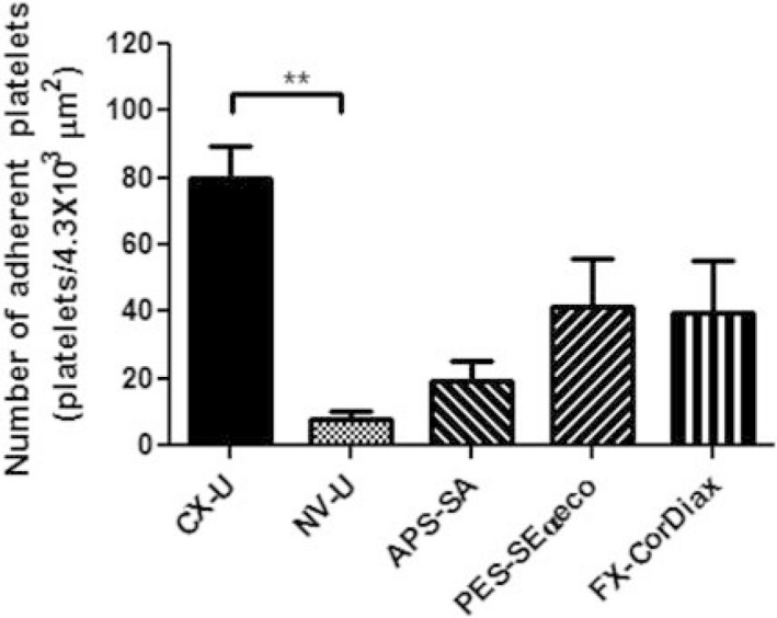
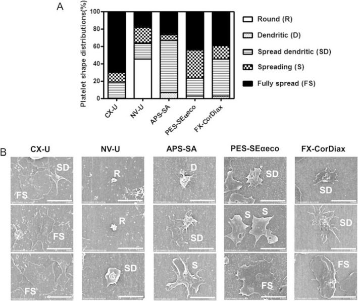
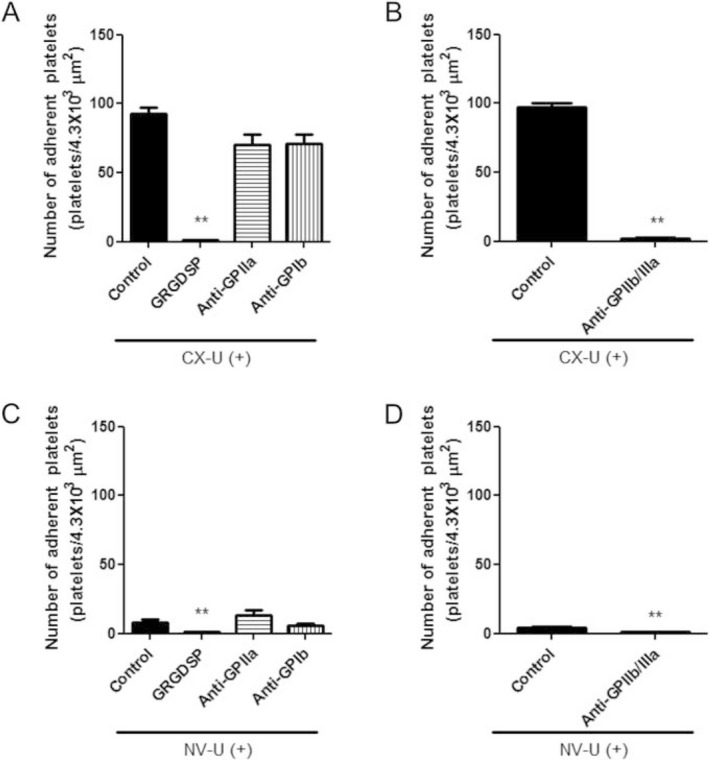
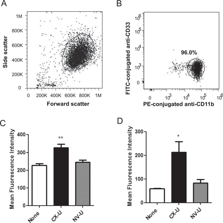
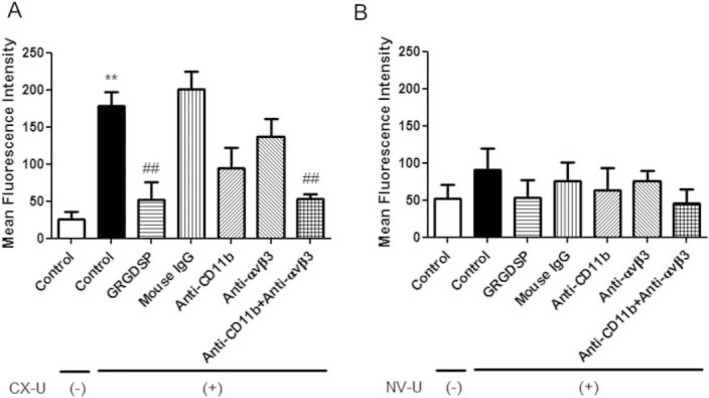
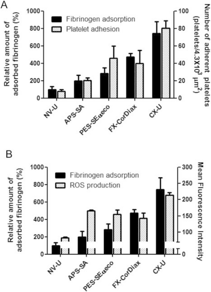
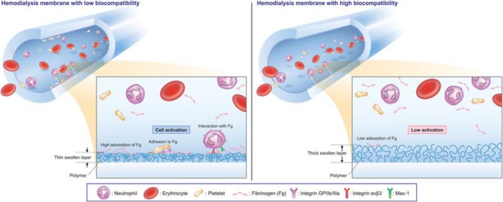
Activation of blood cells during hemodialysis is considered to be a significant determinant of biocompatibility of the hemodialysis membrane because it may affect patient health adversely through microvascular inflammation and oxidative stress. This study found very different cell activation among various polysulfone (PSf) hemodialysis membranes. For example, CX-U, a conventional PSf membrane, induced marked adhesion of platelets to its surface and increased surface expression of activated CD11b and production of reactive oxygen species (ROS) by neutrophils; while NV-U, a hydrophilic polymer-immobilized PSf membrane, caused little platelet adhesion and slight CD11b expression and ROS production by neutrophils. Analysis of the molecular mechanisms of the above phenomena on CX-U and NV-U indicated that anti-integrin GPIIb/IIIa antibody blocked platelet adhesion, and that the combination of anti-CD11b (integrin α subunit of Mac-1) and anti-integrin αvβ3 antibodies blocked ROS production by neutrophils. Plasma-derived fibrinogen, a major ligand of GPIIb/IIIa, Mac-1, and αvβ3 on membranes, was thus analyzed and found to be more adsorbed to CX-U than to NV-U. Moreover, comparison between five PSf membranes showed that the number of adherent platelets and neutrophil ROS production increased with increasing fibrinogen adsorption. These results suggested that fibrinogen, adsorbed on membranes, induced GPIIb/IIIa-mediated platelet activation and Mac-1/αvβ3-mediated neutrophil activation, depending on the amount of adsorption. In conclusion, the use of biocompatible membranes like NV-U, which show lower adsorption of fibrinogen, is expected to reduce hemodialysis-induced inflammation and oxidative stress by minimizing cell activation.

Keywords: Biocompatibility; Fibrinogen; Hemodialysis membrane; Neutrophil; Platelet.


Figures

Figure 1
Figure 1
Number of platelets adherent to the inner surface of hemodialysis (HD) membranes. Data are presented as mean ± SEM of eight independent experiments (using eight different blood donors, respectively). **: P < 0.01 between CX‐U and NV‐U (nonparametric Tukey's multiple comparison).
Figure 2
Figure 2
Morphological changes in platelets adherent to hemodialysis (HD) membranes. A) Platelet shape distributions for different HD membranes. The shape of adherent platelets in the SEM pictures of 5 to 20 fields (enlargement × 5000, area 6.1 × 102 μm2) for each HD membrane was categorized. Each histogram shows the relative percentage of platelets in each of five morphological forms: round or discoid (R), dendritic (D) or early pseudopodial, spread dendritic (SD) or intermediate pseudopodial, spreading (S), and fully spread (FS). (B) Representative scanning electron micrographs of human platelets adherent to different HD membranes are shown (scale bar = 5 μm, enlargement ×10 000).
Figure 3
Figure 3
Effects of integrin antagonists on adhesion of platelets to hemodialysis (HD) membranes.
Figure 4
Figure 4
Activation of neutrophils induced by hemodialysis (HD) membranes in whole blood. The fluorescence intensity of cells was analyzed by flow cytometry. Neutrophils were identified as CD33 low‐positive cells in forward scatter versus log fluorescence of FITC‐conjugated anti‐CD33 dot plot (A). (B) A representative flow cytometry histogram plot showing expression of activated CD11b on neutrophils. Filled histogram, cells with isotype control; solid line, cells without miniMD (None group); dotted line, cells of CX‐U miniMD; dashed line, cells of NV‐U miniMD. (C) Expression of activated CD11b on neutrophils. (D) Granulocytes were gated in forward scatter versus side scatter dot plot. (E) A representative flow cytometry histogram plot showing production of ROS. Filled histogram, autofluorescence of cells; solid line, cells without miniMD (None group); dotted line, cells of CX‐U miniMD; dashed line, cells of NV‐U miniMD. (F) Production of ROS on neutrophils. The ROS production in neutrophils was detected by measuring the intracellular oxidized form of DCFH. Each column represents as mean ± SEM of three independent experiments (using three different blood donors, respectively) *: P < 0.05 versus None (Dunnett's multiple comparison).
Figure 5
Figure 5
Activation of purified neutrophils induced by hemodialysis (HD) membranes. The fluorescence of the neutrophils was measured by flow cytometry. (A) Neutrophils were gated in forward scatter versus side scatter dot plot. (B) The isolated cells were CD33 low positive and CD11b high positive in log fluorescence of FITC‐CD33 versus log fluorescence of PE‐conjugated anti‐CD11b dot plot. The expression of activated CD11b (C) and the intracellular oxidized form of DCFH, an index of ROS production (D) are shown. Data are presented as mean ± SEM of three independent experiments (using three different blood donors, respectively). *: P < 0.05, **: P < 0.01 versus None (Dunnett's or nonparametric Dunnett's multiple comparison).
Figure 6
Figure 6
Effects of integrin antagonists on neutrophil activation induced by CX‐U and NV‐U. Isolated neutrophils suspended in plasma were treated with small pieces of hemodialysis (HD) membrane or without HD membrane (None group) in the absence or presence of indicated antagonists for 30 min at 37°C, and then stained with DCFH‐DA at 37°C for 15 min. The HD membranes used were CX‐U (A) and NV‐U (B). The fluorescence of the neutrophils was measured by flow cytometry to detect the intracellular oxidized form of DCFH, an index of ROS production. Data are presented as mean ± SEM of triplicate measurements from one representative experiment. **: P < 0.01 between Control/CX‐U(‐) versus Control/CX‐U(+) (t‐test), ##: P < 0.01 versus Control/CX‐U(+) (Dunnett's multiple comparison).
Figure 7
Figure 7
Adsorption of fibrinogen onto hemodialysis (HD) membranes and its relationship to cell responses. The left axis and black columns show the relative adsorption of fibrinogen onto HD membranes. Data shows the relative adsorption of fibrinogen onto each HD membrane (%), defining the adsorption of fibrinogen onto NV‐U as 100%. (A) The right axis and dotted columns show the number of platelets adherent to each HD membrane described in Fig. 1. (B) The right axis and dotted columns show the mean fluorescence intensity, which indicates ROS production of neutrophils determined. The fluorescence of the neutrophils was measured by flow cytometry to detect the intracellular oxidized form of DCFH, an index of ROS production. Data for fibrinogen adsorption and ROS production are presented as mean ± SEM of three independent experiments.
Figure 8
Figure 8
Schematic illustration showing the means by which hemodialysis membranes affect platelet and neutrophil activation.

Similar articles

Cited by

References

    1. Daugirdas JT, Bernardo AA. Hemodialysis effect on platelet count and function and hemodialysis‐associated thrombocytopenia. Kidney Int 2012;82:147–57. - PubMed
    1. Sirolli V, Ballone E, Di Stante S, Amoroso L, Bonomini M. Cell activation and cellular‐cellular interactions during hemodialysis: effect of dialyzer membrane. Int J Artif Organs 2002;25:529–37. - PubMed
    1. Yoon JW, Pahl MV, Vaziri ND. Spontaneous leukocyte activation and oxygen‐free radical generation in end‐stage renal disease. Kidney Int 2007;71:167–72. - PubMed
    1. Pertosa G, Grandaliano G, Gesualdo L, Schena FP. Clinical relevance of cytokine production in hemodialysis. Kidney Int Suppl 2000;58:S104–11. - PubMed
    1. Kakuta T, Komaba H, Takagi N, et al. A prospective multicenter randomized controlled study on interleukin‐6 removal and induction by a new hemodialyzer with improved biocompatibility in HD patients: a pilot study. Ther Apher Dial 2016;20:569–78. - PubMed