Review
. 2019 Feb;28(2):292-304.
doi: 10.1002/pro.3509.

Emerging structural biology of lipid G protein-coupled receptors

Affiliations
Review

Emerging structural biology of lipid G protein-coupled receptors

Martin Audet et al. Protein Sci. 2019 Feb.

Abstract

The first crystal structure of a G protein-coupled receptor (GPCR) was that of the bovine rhodopsin, solved in 2000, and is a light receptor within retina rode cells that enables vision by transducing a conformational signal from the light-induced isomerization of retinal covalently bound to the receptor. More than 7 years after this initial discovery and following more than 20 years of technological developments in GPCR expression, stabilization, and crystallography, the high-resolution structure of the adrenaline binding β2 -adrenergic receptor, a ligand diffusible receptor, was discovered. Since then, high-resolution structures of more than 53 unique GPCRs have been determined leading to a significant improvement in our understanding of the basic mechanisms of ligand-binding and ligand-mediated receptor activation that revolutionized the field of structural molecular pharmacology of GPCRs. Recently, several structures of eight unique lipid-binding receptors, one of the most difficult GPCR families to study, have been reported. This review presents the outstanding structural and pharmacological features that have emerged from these new lipid receptor structures. The impact of these findings goes beyond mechanistic insights, providing evidence of the fundamental role of GPCRs in the physiological integration of the lipid signaling system, and highlighting the importance of sustained research into the structural biology of GPCRs for the development of new therapeutics targeting lipid receptors.

Keywords: G protein-coupled receptor; allosteric ligands; bioactive lipids; crystal structures; membrane proteins.


Figures

Figure 1
Figure 1
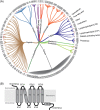
G protein‐coupled receptors (GPCR). (A) Coverage of non‐olfactory class A GPCRs grouped respective to ligand type. The crystallized receptors are indicated by a red dot. Adapted from GPCRdb.9 (B) Schematic of the two‐dimensional topology of a GPCR consists of seven transmembrane helical domains, an extracellular N‐terminus, three extracellular loops (ECLs), three intracellular loops (ICLs), an additional intracellular helical domain that is parallel to the membrane plane followed by an intracellular C‐terminus.
Figure 2
Figure 2
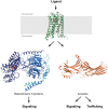
Signaling of GPCRs. Ligand interactions with the receptor's binding site triggers the intracellular engagement of signaling effectors, such as the heterotrimetic G proteins or the arrestins, leading to a downstream intracellular signaling cascades and physiological responses. Cannabinoid receptor 1 (CB1) (green ribbons) and agonist ligand AM11542 (green spheres) are from PDB access code 5xra. Heterotrimeric G protein αi1β1γ2 (blue ribbons and spheres) is from PDB access code 1gp2. Arrestin‐3 (orange ribbons) is from PDB access code 5tv1.
Figure 3
Figure 3
Two‐dimensional structures of important lipid receptor ligands. (A) Bioactive lipids and (B) synthetic derivatives. The ligands are classified according to their reported efficacy. The target receptor is indicated in parenthesis below compound's name.
Figure 4
Figure 4
Overall crystal structures of lipid receptors in complex with ligands. (A and B) Receptors displaying a (A) β‐strand lid or (B) N‐terminal α‐helical lid on the binding site. (C) CB1 shows both features with an α‐helical lid in the agonist‐bound state (see Figure 2) and β‐strand lid in the antagonist‐bound state. Receptors are shown as ribbons and ligands as spheres: prostaglandin E2 receptor 3 (EP3) bound to misoprostol (PDB access code 6m9t; white); GPR40 bound to TAK875 (PDB access code 4phu; blue), apo lysophosphatidic acid receptor 6 (LPA6) (PDB access code 5xsz; yellow), leukotriene B4 receptor 1 (BLT1) bound to BIIL260 (PDB access code 5x33; magenta), sphingosine‐1‐phosphate receptor 1 (S1P1) bound to ML056 (PDB access code 3v2y; green), lysophosphatidic acid receptor 1 (LPA1) bound to ONO9780307 (PDB access code 4z34; purple), platelet‐activating factor receptor (PAFR) bound to ABT491 (PDB access code 5zkq; orange), and CB1 bound to AM6538 (PDB access code 5tgz; brown).
Figure 5
Figure 5
Hypothetical mechanisms of ligand entry into the orthosteric binding sites of lipid receptors. (A) Free diffusing bioactive lipids can access the binding site from the extracellular space and by lateral diffusion in the plane of the membrane. Transporters and enzymes, such as high‐density lipoprotein (HDL) and autotaxin (PDB access code 3nkp), are recruited to the cellular surface and distribute the bioactive lipids in the receptor's local membrane area or directly channel the bioactive lipids into the receptor.45, 49 The crystal structures of lipid receptors suggest a putative ligand entry mechanism in the orthosteric receptor binding site from the classical (B) extracellular side and from the (C) lateral membrane plane of the receptors or (D) both. (E) EP3 receptor in complex with misoprostol show no tunnel that suggests a ligand entry site. The red arrows indicate the putative ligand entry site. The receptors are represented as surfaces and the ligand as spheres. The structures are displayed according to the same color and PDB access code as in Figure 4.
Figure 6
Figure 6
Example of a binding site and activating transition in a lipid receptor. (A) Extracellular view of CB1 receptor in complex with the agonist AM841 (PDB access code 5xr8, active) and the antagonist AM6538 (PDB access code 5tgz, inactive). The inward movement of helices I–II and rotation of helix II reduce the binding site volume in the region formed by helices I, II, and VII. Helix II movement and rotation reposition residues Phe1702.57 and Phe1742.61 to form the agonist binding site floor. Together with the N‐terminal domain that adopts a partial helical conformation, the new binding site configuration is refolded to accommodate only the agonist. (B) Close‐up view of the activating twin microswitch transition implicating residues Phe2003.36 and Trp3566.48. (C) Intracellular view of the CB1 receptor activating the transition. Unlocking the toggle switch residue Trp3566.48 frees the outward movement of helix VI opening the effector coupling intracellular cavity. CB1 receptor bound to the agonist is shown in green cartoon (receptor) and sticks (AM841 ligand). CB1 receptor bound to the antagonist is shown in brown cartoon (receptor) and sticks (AM6538 ligand). (D) Misoprostol tight fit in EP3 receptor orthosteric binding pocket. EP3 receptor is shown as gray surface and misoprostol as light gray spheres. EP3 receptor in complex with misoprostol is from pbdid 6m9t
Figure 7
Figure 7
Allosteric modulation of lipid receptors. (A–C) GPR40 structures in complex with (A) MK8666 (PDB access code 5tzr), (B) compound 1 (PDB access code 5kw2) and (C) both MK8666 and AP8 (PDB access code 5tzy) showing two distinct allosteric sites. (D and E) Overlay of GPR40 in complex with MK8666 (green ribbons and sticks), and Compound 1 (blue ribbons and sticks). The conformational changes between the two structures are indicated by red arrows and dotted lines. Residue Leu1905.46’s induced‐fit allows allosteric ligand AP8 to bind the receptor between the helices V and VI on the outer surface of the seven transmembrane bundle facing the lipid bilayer. Binding of AP8 induces the folding of ICL2, probably favoring the engagement of intracellular effectors.

References

    1. Santos R, Ursu O, Gaulton A, Bento AP, Donadi RS, Bologa CG, Karlsson A, Al‐Lazikani B, Hersey A, Oprea TI, Overington JP (2016) A comprehensive map of molecular drug targets. Nat Rev Drug Discov 16:19. - PMC - PubMed
    1. Sriram K, Insel PA (2018) G protein‐coupled receptors as targets for approved drugs: How many targets and how many drugs? Mol Pharmacol 93:251–258. - PMC - PubMed
    1. Alexander SP, Christopoulos A, Davenport AP, Kelly E, Marrion NV, Peters JA, Faccenda E, Harding SD, Pawson AJ, Sharman JL, Southan C, Davies JA, Collaborators CGTP (2017) The Concise Guide to Pharmacology 2017/18: G protein‐coupled receptors. Br J Pharmacol 174:S17–S129. - PMC - PubMed
    1. Palczewski K, Kumasaka T, Hori T, Behnke CA, Motoshima H, Fox BA, Le Trong I, Teller DC, Okada T, Stenkamp RE, Yamamoto M, Miyano M (2000) Crystal structure of rhodopsin: A G protein‐coupled receptor. Science 289:739–745. - PubMed
    1. Cherezov V, Rosenbaum DM, Hanson MA, Rasmussen SG, Thian FS, Kobilka TS, Choi HJ, Kuhn P, Weis WI, Kobilka BK, Stevens RC (2007) High‐resolution crystal structure of an engineered human beta 2‐adrenergic G protein‐coupled receptor. Science 318:1258–1265. - PMC - PubMed

MeSH terms

Substances

LinkOut - more resources