. 2019 May;10(3):738-744.
doi: 10.1111/jdi.12934. Epub 2018 Oct 13.

Pathological significance of urinary complement activation in diabetic nephropathy: A full view from the development of the disease

Affiliations

Pathological significance of urinary complement activation in diabetic nephropathy: A full view from the development of the disease

Jing-Min Zheng et al. J Diabetes Investig. 2019 May.

Abstract

Aims/introduction: The aim of the present study was to obtain a full view of the changes of urinary complement activation products in the development of diabetic nephropathy and explore their possible significance in the disease process.

Materials and methods: A total of 62 patients at different stages of diabetic nephropathy, 20 diabetes patients without nephropathy and 20 healthy persons were enrolled. Urinary complement activation products, including C3a, C5a and C5b-9, were measured, and their associations with the progression of the disease were analyzed.

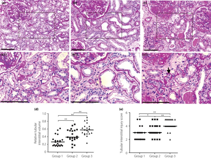
Results: The urinary complement activation products increased markedly since the proteinuria stage, and were parallel with the progression of diabetic nephropathy. More severe renal tubular damage was observed in patients with higher levels of urinary complement activation products. The urinary complement activation products levels correlated closely with renal tubulointerstitial injury score and relative tubular interstitial volume. Multivariate regression analysis showed that elevated urinary complement activation products were independent risk factors for tubular injury in diabetic nephropathy patients.

Conclusions: Urinary complement activation might have a role in renal tubular interstitial injury in patients with diabetic nephropathy, especially in patients at a later stage of the disease.

Keywords: Diabetic nephropathy; Tubular injury; Urinary complement activation products.


Figures

Figure 1
Figure 1
Urinary levels of C3a, C5a and C5b‐9 in patients at different clinical stages of diabetic nephropathy. As the data about the levels of urinary complement activation products (including C3a, C5a and C5b‐9) were determined to be non‐normal, multigroup comparison was carried out using the Kruskal–Wallis test. The difference between the two groups was compared with a non‐parametric Wilcoxon rank sum test. (a) Urinary levels of C3a in different groups. (b) Urinary levels of C5a in different groups. (c) Urinary levels of C5b‐9 in different groups. Thick black line shows the median. **< 0.01. DMG, diabetes mellitus group (without nephropathy); MG, microalbuminuria stage group; NG, normal control group; PG, proteinuria stage group; RIG, renal insufficiency stage group.
Figure 2
Figure 2
More severe renal tubular damage, such as a higher proportion of atrophic tubules, larger tubular interstitial area, more severe degree of brush border loss and more tubular epithelial cells detached from the tubular basement membrane (shown by black arrow), was observed in the renal biopsies of patients with higher levels of urinary complement activation products. According to the level of urinary C3a, all the patients were divided into three groups: group 1, patients with the urinary C3a level within the first third (<9.2 ng/mg·Cr); group 2, patients with the urinary C3a level within the second third (≥9.2 and <893 ng/mg·Cr); group 3, patients with a urinary C3a level within the last third (≥893 ng/mg·Cr). As the data about tubular interstitial injury score and relative interstitial volume were determined to be non‐normal, multigroup comparison was carried out using the Kruskal–Wallis test. The difference between the two groups was compared with a non‐parametric Wilcoxon rank sum test. (a) A representative picture of periodic acid–Schiff staining from a patient in group 1. (b) A representative picture of periodic acid–Schiff staining from a patient in group 2. (c) A representative picture of periodic acid–Schiff staining from a patient in group 3. (d) The relative tubular interstitial volume in different groups. (e) The tubular interstitial injury score in different groups. (a1,b1,c1) Partial enlargement of (a–c), respectively. Scale bars (a–c, a1–c1), 50 μM. Thick black line in (d) and (e) shows the median.*< 0.05; **< 0.01.

Similar articles

Cited by

References

    1. Molitch ME, Adler AI, Flyvbjerg A, et al Diabetic kidney disease: a clinical update from Kidney Disease: improving global outcomes. Kidney Int 2015; 87: 20–30. - PMC - PubMed
    1. Lim AK. Diabetic nephropathy ‐ complications and treatment. Int J Nephrol Renovasc Dis 2014; 7: 361–381. - PMC - PubMed
    1. Fried LF, Emanuele N, Zhang JH, et al Combined angiotensin inhibition for the treatment of diabetic nephropathy. N Engl J Med 2013; 369: 1892–1903. - PubMed
    1. de Zeeuw D, Akizawa T, Audhya P, et al Bardoxolone methyl in type 2 diabetes and stage 4 chronic kidney disease. N Engl J Med 2013; 369: 2492–2503. - PMC - PubMed
    1. Flyvbjerg A. The role of the complement system in diabetic nephropathy. Nat Rev Nephrol 2017; 13: 311–318. - PubMed

MeSH terms