

High-fat diet-induced hypertension is associated with a proinflammatory T cell profile in male and female Dahl salt-sensitive rats
- PMID: 30239234
- PMCID: PMC6336972
- DOI: 10.1152/ajpheart.00389.2018
High-fat diet-induced hypertension is associated with a proinflammatory T cell profile in male and female Dahl salt-sensitive rats
Abstract
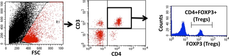
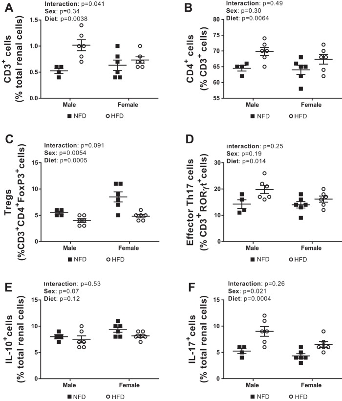
Evidence supports a sex difference in the impact of a high-fat diet (HFD) on cardiovascular outcomes, with male experimental animals exhibiting greater increases in blood pressure (BP) than female experimental animals. The immune system has been implicated in HFD-induced increases in BP, and there is a sex difference in T-cell activation in hypertension. The goal of this study was to determine the impact of HFD on BP and aortic and renal T cell profiles in male and female Dahl salt-sensitive (DSS) rats. We hypothesized that male DSS rats would have greater increases in BP and T cell infiltration in response to a HFD compared with female DSS rats. BP was measured by tail-cuff plethysmography, and aortic and renal T cells were assessed by flow cytometric analysis in male and female DSS rats on a normal-fat diet (NFD) or HFD from 12 to 16 wk of age. Four weeks of HFD increased BP in male and female DSS rats to a similar degree. Increases in BP were accompanied by increased percentages of CD4+ T cells and T helper (Th)17 cells in both sexes, although male rats had more proinflammatory T cells. Percentages of renal CD3+ and CD4+ T cells as well as Th17 cells were increased in both sexes by the HFD, although the increase in CD3+ T cells was greater in male rats. HFD also decreased the percentage of aortic and renal regulatory T cells in both sexes, although female rats maintained more regulatory T cells than male rats regardless of diet. In conclusion, both male and female DSS rats exhibit BP sensitivity to a HFD; however, the mechanisms mediating HFD-induced increases in BP may be distinct as male rats exhibit greater increases in the percentage of proinflammatory T cells than female rats. NEW & NOTEWORTHY Our study demonstrates that male and female Dahl salt-sensitive rats exhibit similar increases in blood pressure to a high-fat diet and an increase in aortic and renal T cells. These results are in contrast to studies showing that female rats remain normotensive and/or upregulate regulatory T cells in response to hypertensive stimuli compared with male rats. Our data suggest that a 4-wk high-fat diet has sex-specific effects on the T cell profile in Dahl salt-sensitive rats.
Keywords: T cells; aorta; inflammation; kidney; sex differences.
Figures


Similar articles
-
T cell knockout attenuates HFD-induced increases in blood pressure in female and male Dahl rats.Clin Sci (Lond). 2025 Sep 9;139(17):941-954. doi: 10.1042/CS20257273. Clin Sci (Lond). 2025. PMID: 40810662
-
Sex Differences in Renal Inflammation and Injury in High-Fat Diet-Fed Dahl Salt-Sensitive Rats.Hypertension. 2018 Nov;72(5):e43-e52. doi: 10.1161/HYPERTENSIONAHA.118.11485. Hypertension. 2018. PMID: 30354819 Free PMC article.
-
Effects of high-fat diet on sympathetic neurotransmission in mesenteric arteries from Dahl salt-sensitive rat.Auton Neurosci. 2019 Dec;222:102599. doi: 10.1016/j.autneu.2019.102599. Epub 2019 Oct 31. Auton Neurosci. 2019. PMID: 31731103 Free PMC article.
-
Sex differences in T cells in hypertension.Clin Ther. 2014 Dec 1;36(12):1882-1900. doi: 10.1016/j.clinthera.2014.07.011. Epub 2014 Aug 16. Clin Ther. 2014. PMID: 25134971 Free PMC article. Review.
-
Role of Female Sex Hormones and Immune Response in Salt-Sensitive Hypertension Development: Evidence from Experimental Models.Curr Hypertens Rep. 2023 Nov;25(11):405-419. doi: 10.1007/s11906-023-01257-1. Epub 2023 Sep 7. Curr Hypertens Rep. 2023. PMID: 37676461 Review.
Cited by
-
Transfer of Th17 from Adult Spontaneous Hypertensive Rats Accelerates Development of Hypertension in Juvenile Spontaneous Hypertensive Rats.Biomed Res Int. 2021 Feb 20;2021:6633825. doi: 10.1155/2021/6633825. eCollection 2021. Biomed Res Int. 2021. PMID: 33688497 Free PMC article.
-
Activated pathogenic Th17 lymphocytes induce hypertension following high-fructose intake in Dahl salt-sensitive but not Dahl salt-resistant rats.Dis Model Mech. 2020 May 27;13(5):dmm044107. doi: 10.1242/dmm.044107. Dis Model Mech. 2020. PMID: 32179549 Free PMC article.
-
The Vascular Consequences of Metabolic Syndrome: Rodent Models, Endothelial Dysfunction, and Current Therapies.Front Pharmacol. 2020 Mar 4;11:148. doi: 10.3389/fphar.2020.00148. eCollection 2020. Front Pharmacol. 2020. PMID: 32194403 Free PMC article. Review.
-
Luteolin Relieves Metabolic Dysfunction-Associated Fatty Liver Disease Caused by a High-Fat Diet in Rats Through Modulating the AdipoR1/AMPK/PPARγ Signaling Pathway.Int J Mol Sci. 2025 Apr 17;26(8):3804. doi: 10.3390/ijms26083804. Int J Mol Sci. 2025. PMID: 40332475 Free PMC article.
-
PDZ-Binding Kinase, a Novel Regulator of Vascular Remodeling in Pulmonary Arterial Hypertension.Circulation. 2024 Jul 30;150(5):393-410. doi: 10.1161/CIRCULATIONAHA.123.067095. Epub 2024 Apr 29. Circulation. 2024. PMID: 38682326 Free PMC article.
References
-
- Bean C, Spencer SK, Bowles T, Kyle PB, Williams JM, Gibbens J, Wallace K. Inhibition of T-cell activation attenuates hypertension, TNFα, IL-17, and blood-brain barrier permeability in pregnant rats with angiogenic imbalance. Am J Reprod Immunol 76: 272–279, 2016. doi:10.1111/aji.12547. - DOI - PMC - PubMed
Publication types
MeSH terms
Grants and funding
LinkOut - more resources
Full Text Sources
Other Literature Sources
Medical
Research Materials