. 2018 Nov 2;46(19):10286-10301.
doi: 10.1093/nar/gky838.

Structural insights into the mechanism of double strand break formation by Hermes, a hAT family eukaryotic DNA transposase

Affiliations

Structural insights into the mechanism of double strand break formation by Hermes, a hAT family eukaryotic DNA transposase

Alison B Hickman et al. Nucleic Acids Res. .

Abstract

Some DNA transposons relocate from one genomic location to another using a mechanism that involves generating double-strand breaks at their transposon ends by forming hairpins on flanking DNA. The same double-strand break mode is employed by the V(D)J recombinase at signal-end/coding-end junctions during the generation of antibody diversity. How flanking hairpins are formed during DNA transposition has remained elusive. Here, we describe several co-crystal structures of the Hermes transposase bound to DNA that mimics the reaction step immediately prior to hairpin formation. Our results reveal a large DNA conformational change between the initial cleavage step and subsequent hairpin formation that changes which strand is acted upon by a single active site. We observed that two factors affect the conformational change: the complement of divalent metal ions bound by the catalytically essential DDE residues, and the identity of the -2 flanking base pair. Our data also provides a mechanistic link between the efficiency of hairpin formation (an A:T basepair is favored at the -2 position) and Hermes' strong target site preference. Furthermore, we have established that the histidine residue within a conserved C/DxxH motif present in many transposase families interacts directly with the scissile phosphate, suggesting a crucial role in catalysis.


Figures

Figure 1.
Figure 1.
(A) Overview of cut-and-paste DNA transposition. Transposases catalyze the excision of a transposon (in blue) from one genomic location by introducing double-strand breaks at each end. Excised transposons are then integrated into a new genomic site, a process usually accompanied by the generation of short flanking target site duplications (TSDs). (B) Comparison of the steps comprising transposition when a hairpin is formed on flanking DNA (left, hAT transposons or during V(D)J recombination) or on the transposon end (right, IS4 family of insertion sequences and eukaryotic piggyBac element). In step (i), hAT transposases cleave the non-transferred strand (NTS) one nt into the flank whereas the V(D)J recombinase cleaves precisely at the coding end-recombination signal sequence junction; TS: transferred strand. Throughout, flanking DNA is shown in green and transposon DNA in blue. (C) Oligonucleotides used for crystallography, ‘DNA(C:G)’ on top and ‘DNA(A:T)’ on the bottom. Also shown is the preferred target site for integration of Hermes.
Figure 2.
Figure 2.
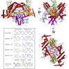
Structure of Hermes/DNA complex, here the DNA(A:T) complex bound to Mn2+. Hermes coloring corresponds to its domains: dimerization domain in purple, insertion domain in red, and the RNase H-like catalytic domain in orange. Inset: Comparison of DNA conformations observed in four Hermes-DNA structures prior to hairpin formation to (top) ideal B-form DNA of the same bp length and (bottom) the nicked paired-end complex of RAG1/2 (22). In all cases, the trajectory of the free 5′-end of the transposon end is away from the active site.
Figure 3.
Figure 3.
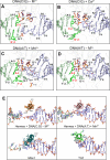
(A–D) Comparison of active site regions of four Hermes-DNA structures prior to hairpin formation. Stick colors correspond to the protein domain and DNA colors in Figure 1. The variable bp at –2 is colored bright green, and the scissile phosphate is indicated in orange. The distance between the nucleophilic 3′-OH group and the scissile phosphate is marked. Note that in the complex of Hermes with DNA(C:G) in the absence of metal ions (A), the second nt on the NTS (i.e. A2) is disordered and not visible in the electron density. (E) The interaction between the α-helix bearing the third catalytic acidic residue and the transposon tip. In the DNA(C:G) complex structure in the absence of metal ions (upper left), the catalytic helix (residues 569–581) does not interact with the DNA. Once the DNA conformational change has occurred, the helix is inserted into the minor groove close to the transposon tip (as shown here for the DNA(A:T) structure). The same insertion is observed for Mos1 (4U7B; residues 281–297 are shown) and Tn5 (1MUH; residues 323–334 are shown) transposases bound to their transposon ends.
Figure 4.
Figure 4.
Metal ion binding sites. (A) Complex between Hermes and DNA(C:G) after soaking in Ca2+. (B) Active site of the complex between Hermes and DNA(A:T) after soaking in Mn2+. (C) Third Mn2+ ion binding site in the complex between Hermes and DNA(A:T) after soaking in Mn2+. (D) Active site of the complex between Hermes and DNA(A:T) without metal ion soaking. Water molecules are shown as red spheres.
Figure 5.
Figure 5.
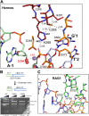
(A) A network of interactions link C265, H268, and the scissile phosphate prior to hairpin formation. Shown is HermesΔ complexed with DNA(A:T) in the presence of Mn2+. (B) In vitro hairpin formation requires H268 of the conserved CxxH motif. The hairpin assay using full-length protein and the oligonucleotide shown on the left was performed as a function of time under conditions as described in Materials and Methods. Reaction products were run on a 20% acrylamide TBE-urea gel and detection was by silver staining. (C) The active site of RAG1 prior to hairpin formation, shown for mouse RAG1/2 (PDB ID: 5ZE1).
Figure 6.
Figure 6.
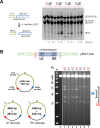
(A) In vitro hairpin formation as a function of the –2 bp. The reaction was run as a function of time using HermesΔ and the oligonucleotide shown on the left. Reaction products were run on a 20% acrylamide TBE-urea gel and detection was by silver staining. (B) In vitro end cleavage as a function of changing the –2 bp on the left end (LE) and right end (RE) of a Hermes minitransposon in the plasmid pRX1. In each lane, the –2 bp on the LE of the Hermes minitransposon is indicated at the top in red and that on the RE is indicated in blue. The assay was performed under conditions as described in Materials and Methods.
Figure 7.
Figure 7.
Proposed ‘ping-pong’ mechanism of Hermes transposition. The active site is used first in one configuration and then in the reverse direction, with the attacking nucleophile and the leaving group alternating sides of a centrally bound scissile phosphate group. The scissile phosphate, presumed to undergo stereochemical inversion with each step, is marked by the orange circle.

References

    1. Rankin D.J., Rocha E.P.C., Brown S.P.. What traits are carried on mobile genetic elements, and why. Heredity. 2011; 106:1–10. - PMC - PubMed
    1. Gillings M.R., Paulsen I.T., Tetu S.G.. Genomics and the evolution of antibiotic resistance. Ann. N. Y. Acad. Sci. 2017; 1388:92–107. - PubMed
    1. Chuong E.B., Elde N.C., Feschotte C.. Regulatory activities of transposable elements: from conflicts to benefits. Nat. Rev. Genet. 2017; 18:71–86. - PMC - PubMed
    1. Siguier P., Gourbeyre E., Chandler M.. Bacterial insertion sequences: their genomic impact and diversity. FEMS Microbiol. Rev. 2014; 38:865–891. - PMC - PubMed
    1. Hickman A.B., Dyda F.. DNA transposition at work. Chem. Rev. 2016; 116:12758–12784. - PMC - PubMed

Publication types

LinkOut - more resources