. 2018 Nov 1;77(11):1039-1054.
doi: 10.1093/jnen/nly085.

A Revised Diagnostic Classification of Canine Glioma: Towards Validation of the Canine Glioma Patient as a Naturally Occurring Preclinical Model for Human Glioma

Affiliations

A Revised Diagnostic Classification of Canine Glioma: Towards Validation of the Canine Glioma Patient as a Naturally Occurring Preclinical Model for Human Glioma

Jennifer W Koehler et al. J Neuropathol Exp Neurol. .

Abstract

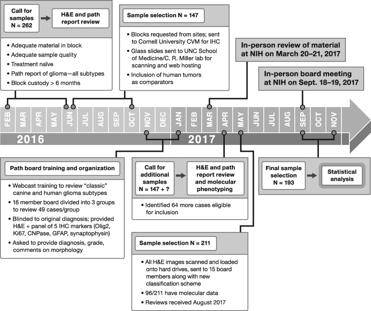
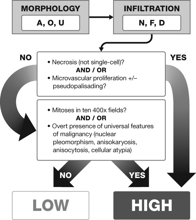
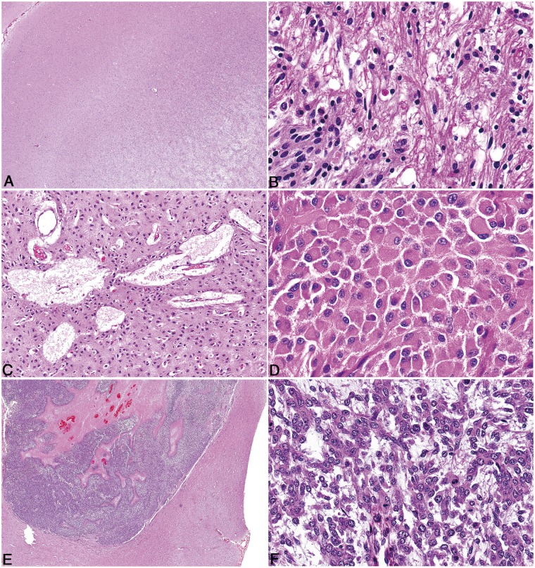
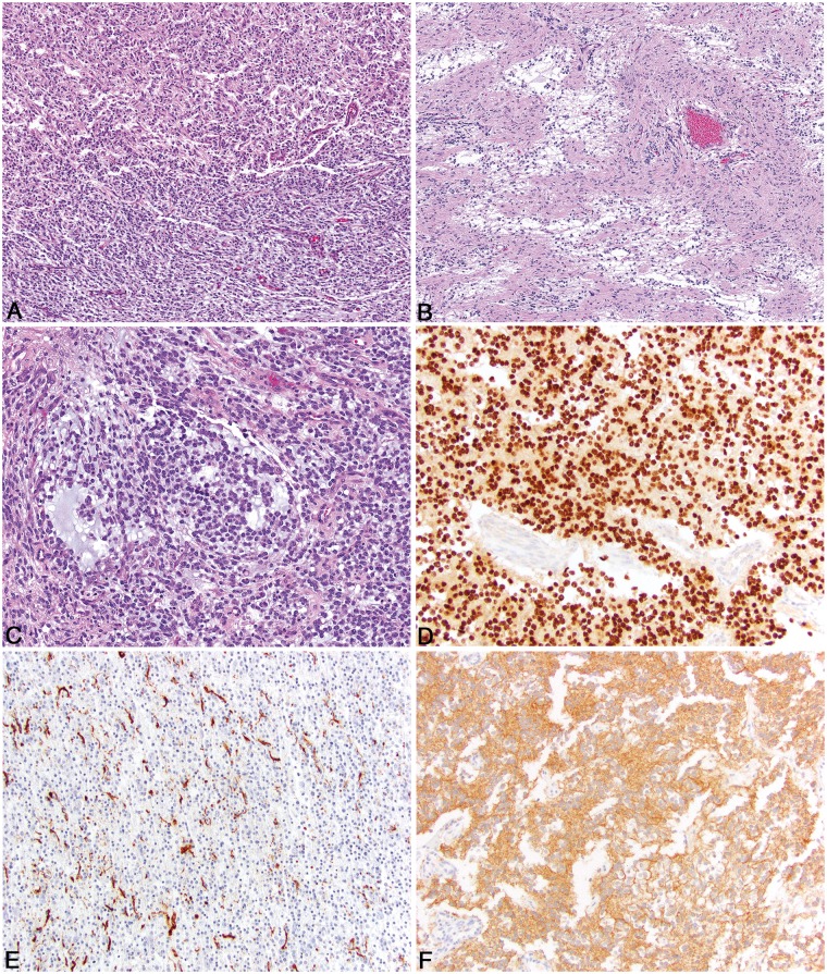
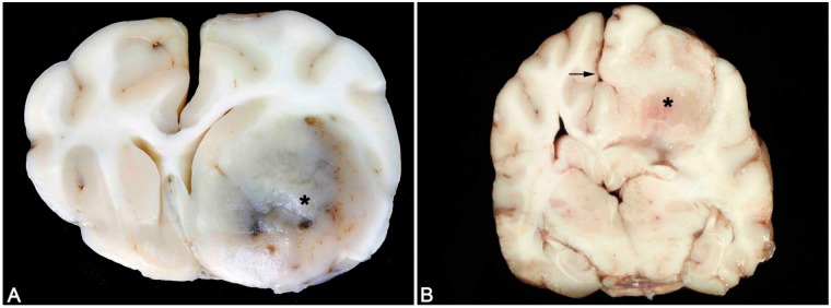
The National Cancer Institute-led multidisciplinary Comparative Brain Tumor Consortium (CBTC) convened a glioma pathology board, comprising both veterinarian and physician neuropathologists, and conducted a comprehensive review of 193 cases of canine glioma. The immediate goal was to improve existing glioma classification methods through creation of a histologic atlas of features, thus yielding greater harmonization of phenotypic characterization. The long-term goal was to support future incorporation of clinical outcomes and genomic data into proposed simplified diagnostic schema, so as to further bridge the worlds of veterinary and physician neuropathology and strengthen validity of the dog as a naturally occurring, translationally relevant animal model of human glioma. All cases were morphologically reclassified according to a new schema devised by the entire board, yielding a majority opinion diagnosis of astrocytoma (43, 22.3%), 19 of which were low-grade and 24 high-grade, and oligodendroglioma (134, 69.4%), 35 of which were low-grade and 99 were high-grade. Sixteen cases (8.3%) could not be classified as oligodendroglioma or astrocytoma based on morphology alone and were designated as undefined gliomas. The simplified classification scheme proposed herein provides a tractable means for future addition of molecular data, and also serves to highlight histologic similarities and differences between human and canine glioma.


Figures

FIGURE 1.
FIGURE 1.
Timeline of events and workflow for the Comparative Brain Tumor Consortium glioma pathology board.
FIGURE 2.
FIGURE 2.
Decision-tree approach for the reclassification of 193 canine gliomas assessed by the Comparative Brain Tumor Consortium glioma pathology board. A = astrocytic morphology; O = oligodendrogial morphology; U = undefined morphology; N = no infiltration; F = focal infiltration; D = diffuse infiltration.
FIGURE 3.
FIGURE 3.
Pathologic features of canine oligodendroglioma. (A) High-grade oligodendroglioma in the cerebrum with effacement of the grey matter and minimal invasion of the subjacent white matter, despite the presence of multiple hallmarks of high-grade such as necrosis and microvascular proliferation. (B) High-grade oligodendroglioma with extensive intraventricular spread (Asterisk = ventricular lumen). (C) High-grade oligodendroglioma with extensive subpial and meningeal spread. (D) Well-differentiated oligodendrogliomas have moderate to dense cellularity, and often have processing-induced retraction of cytoplasm resulting in a “honeycomb” or “fried egg” appearance. (E) Features of poor fixation in canine oligodendroglioma include pyknotic nuclei, poor nuclear detail, tissue discohesion, tinctorial abnormalities, and poor stain uptake in intravascular erythrocytes. (F) Vascular proliferation that comprises increased numbers of congested, branching, and occasionally tortuous vessels lined by a single layer of hypertrophied endothelium, does not meet the criteria for true microvascular proliferation.
FIGURE 4.
FIGURE 4.
Pathologic features of canine oligodendroglioma. (A) In high-grade oligodendroglioma, cells with large, deeply cleaved, folded, or irregular nuclei (arrows) with occasionally pronounced nuclear molding are present. (B) High-grade oligodendrogliomas contain variably robust peripheral foci of microvascular proliferation characterized by tortuous clusters of vessels lined by multiple layers of endothelial cells and pericytes. (C) High-grade oligodendrogliomas have areas of necrosis which in some cases have cellular palisading (arrow) along the margin of the necrotic area. (D) In high-grade oligodendrogliomas, not all regions of necrosis are rimmed by palisading neoplastic cells. (E) In some oligodendrogliomas that have otherwise typical morphology, neoplastic cells are arranged in a pattern that mimics that of ependymal pseudorosettes. (F) Oligodendrogliomas frequently contain moderate to abundant amounts of basophilic, myxoid matrix.
FIGURE 5.
FIGURE 5.
Pathologic features of canine astrocytoma. (A) Low-grade astrocytomas are typically invasive, variably cellular, and distorted the parenchyma. (B) The nuclei of neoplastic astrocytes are typically ovoid to elongate and had a wide range in size with a finely stippled chromatin pattern and 1–2 small nucleoli. (C) Multifocal, but scattered microcysts containing degenerate, proteinaceous material or mucin are present in some astrocytomas. (D) Gemistocyte-like cell morphology in a low-grade gemistocytic astrocytoma. (E) High-grade astrocytomas have large regions of geographic necrosis, with or without palisading along the areas of necrosis. (F) In some astrocytomas, interlacing fascicles of spindle cells predominate in the neoplasm.
FIGURE 6.
FIGURE 6.
Pathologic features of undefined canine glioma and immunohistochemical features. (A) A roughly equal proportion of oligodendroglioma-like morphology (top) and astrocytoma-like morphology bottom). (B) Two distinct populations of neoplastic cells with small, basophilic cells admixed with mucin separated by islands of less dense neoplastic cells that are more astrocytic in morphology. (C) Regions of basophilic mucin deposition in an undefined canine glioma. (D) Olig2 immunoreactivity in canine oligodendroglioma is characterized by diffuse, intense intranuclear immunoreactivity in virtually all of the neoplastic cells. (E) GFAP immunoreactivity in canine astrocytoma is often patchy, but intense cytoplasmic immunoreactivity is noted in approximately 30% of the neoplastic cells. (F) CNPase immunoreactivity in canine oligodendroglioma is diffusely cytoplasmic and specific for the neoplastic cells.
FIGURE 7.
FIGURE 7.
Typical gross appearances of canine oligodendroglioma and astrocytoma. (A) Canine oligodendroglioma. A well-demarcated, gelatinous gray mass is present in the region of the ventral frontal cortex and basal ganglia (asterisk) associated with areas of hemorrhage and a midline shift. (B) Canine astrocytoma. A poorly recognizable mass (asterisk) that is of similar color and consistency with the adjacent parenchyma and is associated with a midline shift (arrow).

References

    1. LeBlanc AK, Breen M, Choyke P, et al. Filling in the gaps for man’s best friend: Perspectives from a National Academies Institute of Medicine Workshop on Comparative Oncology. Sci Transl Med 2016;8: 324ps5 - PMC - PubMed
    1. Paoloni M, Khanna C.. Translation of new cancer treatments from pet dogs to humans. Nat Rev Cancer 2008;8:147–56 - PubMed
    1. Rowell JL, McCarthy DO, Alvarez CE.. Dog models of naturally occurring cancer. Trends Mol Med 2011;17:380–8 - PMC - PubMed
    1. Khanna C, London C, Vail D, et al. Guiding the optimal translation of new cancer treatments from canine to human cancer patients. Clin Cancer Res 2009;15:5671–7 - PMC - PubMed
    1. Vail DM, MacEwen EG.. Spontaneously occurring tumors of companion animals as models for human cancer. Cancer Invest 2000;18:781–92 - PubMed

Publication types

MeSH terms

Substances