

Multiparity affects conduction properties of pelvic floor nerves in rabbits
- PMID: 30240150
- PMCID: PMC6192397
- DOI: 10.1002/brb3.1105
Multiparity affects conduction properties of pelvic floor nerves in rabbits
Abstract
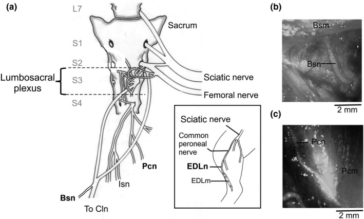
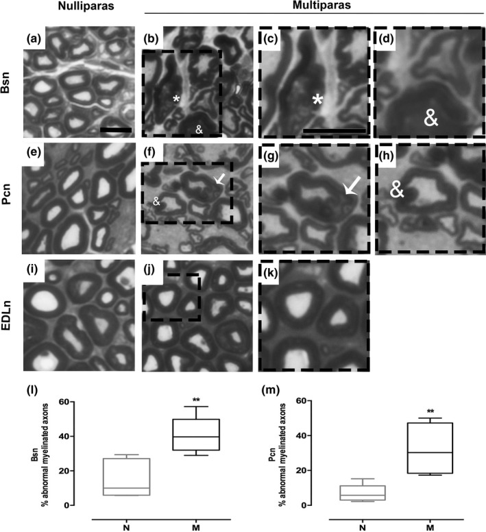
Introduction: Women often develop pelvic floor dysfunction due to damage to the pelvic musculature during childbirth; however, the effect on pelvic floor nerves function is less understood. This study used adult rabbits to evaluate the electrophysiological and histological characteristics of the bulbospongiosus (Bsn) and pubococcygeus nerves (Pcn) in multiparity.
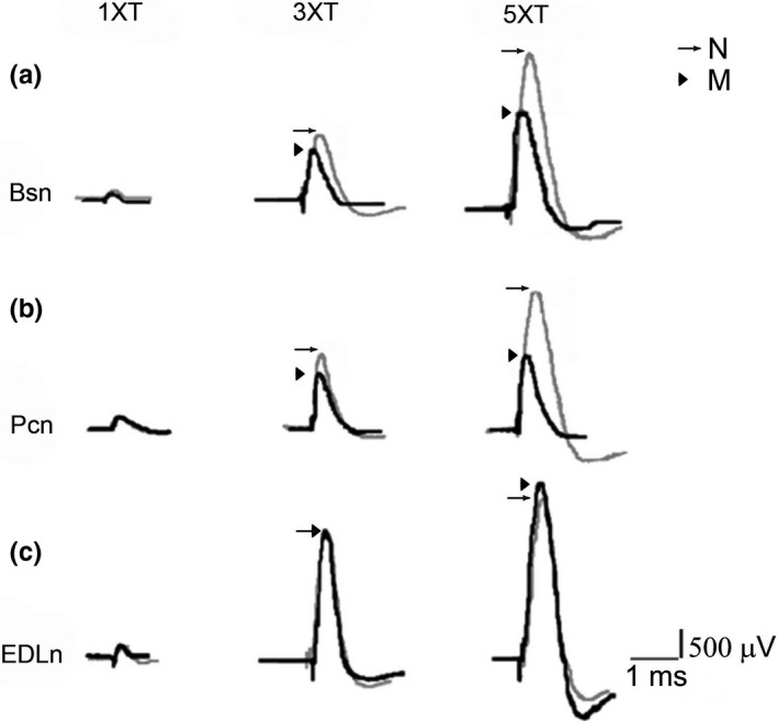
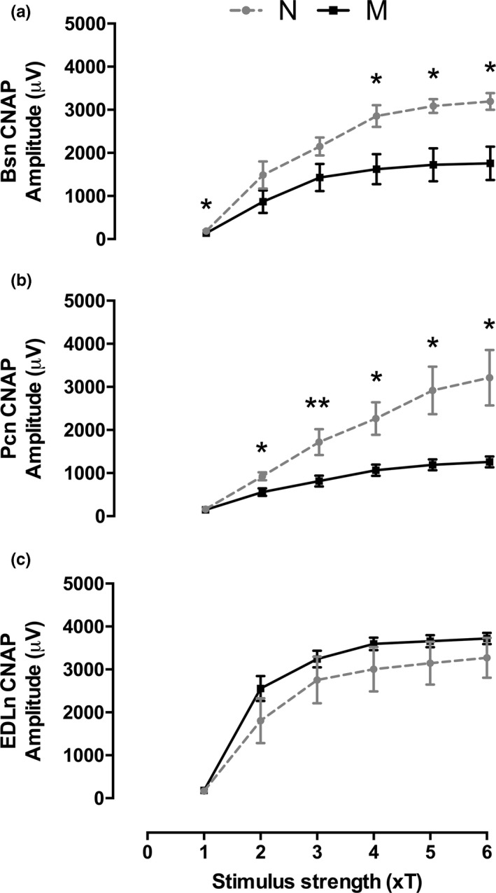
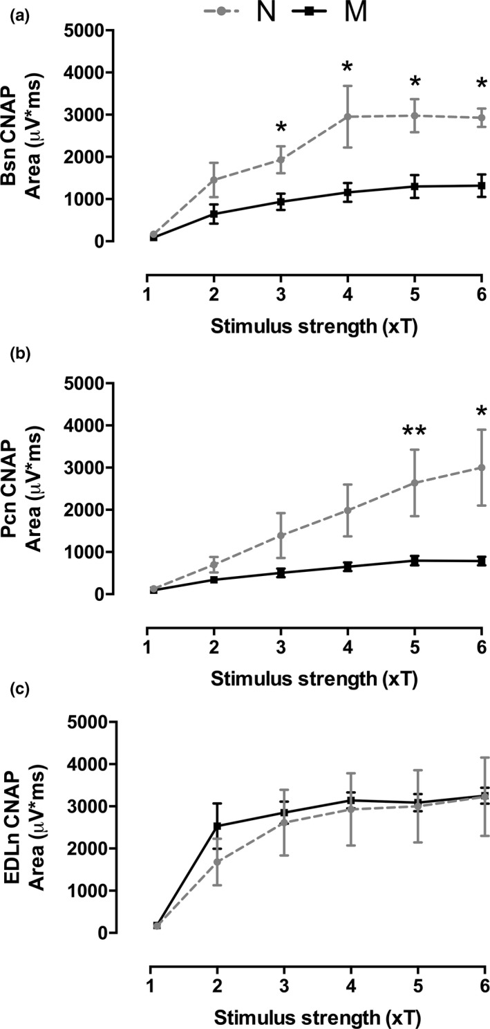
Methods: Compound nerve action potentials (CNAP) were compared between age-matched nulliparous and multiparous animals and associated to the histological characteristics of myelinated axons from the Bsn and Pcn nerves. The extensor digitorum longus nerve (EDLn) was used as negative control. Data were analyzed with unpaired two-tailed Student's t test or Mann-Whitney U test to determine significant differences between groups.
Results: The onset and peak latencies, duration, and conduction velocity of the motor fibers in these pelvic nerves were not significantly different between nulliparous and multiparous animals. However, the peak-to-peak amplitude and area of the CNAP in both Bsn and Pcn were reduced in multiparous rabbits. Histology showed a higher percentage of axons with myelin disorganization caused by multiparity in these pelvic nerves. Together, the data indicate a reduction in the number of functional pelvic axons due to multiparity. As expected, no effect of parity was observed in the EDLn controls.
Conclusions: Present findings demonstrated that multiparity affects myelination and consequently conduction properties in the small pelvic floor nerves.
Keywords: bulbospongiosus muscle; micturition; myelin; pubococcygeus muscle; reproduction; urinary incontinence.
© 2018 The Authors. Brain and Behavior published by Wiley Periodicals, Inc.
Figures
Similar articles
-
Targeted neuromodulation of pelvic floor nerves in aging and multiparous rabbits improves continence.Sci Rep. 2021 May 19;11(1):10615. doi: 10.1038/s41598-021-90088-8. Sci Rep. 2021. PMID: 34011938 Free PMC article.
-
Bladder and urethral dysfunction in multiparous and mature rabbits correlates with abnormal activity of pubococcygeus and bulbospongiosus muscles.Neurourol Urodyn. 2020 Jan;39(1):116-124. doi: 10.1002/nau.24176. Epub 2019 Oct 2. Neurourol Urodyn. 2020. PMID: 31578766 Free PMC article.
-
Fiber type composition of pubococcygeus and bulbospongiosus striated muscles is modified by multiparity in the rabbit.Neurourol Urodyn. 2017 Aug;36(6):1456-1463. doi: 10.1002/nau.23143. Epub 2016 Sep 27. Neurourol Urodyn. 2017. PMID: 27677101
-
[Neurophysiological evaluation of the pelvic floor].Rev Neurol. 1998 Mar;26(151):432-8. Rev Neurol. 1998. PMID: 9585958 Review. Spanish.
-
Postpartum pelvic floor trauma.Curr Opin Obstet Gynecol. 2009 Dec;21(6):474-9. doi: 10.1097/GCO.0b013e328332a84e. Curr Opin Obstet Gynecol. 2009. PMID: 19855276 Review.
Cited by
-
Hybrid Bionic Nerve Interface for Application in Bionic Limbs.Adv Sci (Weinh). 2023 Dec;10(35):e2303728. doi: 10.1002/advs.202303728. Epub 2023 Oct 15. Adv Sci (Weinh). 2023. PMID: 37840396 Free PMC article.
-
Targeted neuromodulation of pelvic floor nerves in aging and multiparous rabbits improves continence.Sci Rep. 2021 May 19;11(1):10615. doi: 10.1038/s41598-021-90088-8. Sci Rep. 2021. PMID: 34011938 Free PMC article.
-
Signs of damage in pelvic floor muscles at the end of pregnancy in rabbits.Int Urogynecol J. 2019 Jun;30(6):977-984. doi: 10.1007/s00192-019-03872-6. Epub 2019 Feb 1. Int Urogynecol J. 2019. PMID: 30706078
-
Bladder and urethral dysfunction in multiparous and mature rabbits correlates with abnormal activity of pubococcygeus and bulbospongiosus muscles.Neurourol Urodyn. 2020 Jan;39(1):116-124. doi: 10.1002/nau.24176. Epub 2019 Oct 2. Neurourol Urodyn. 2020. PMID: 31578766 Free PMC article.
-
Neuromodulation Improves Stress Urinary Incontinence-Like Deficits in Female Rabbits.IEEE Open J Eng Med Biol. 2024 Jun 3;6:10-19. doi: 10.1109/OJEMB.2024.3408454. eCollection 2025. IEEE Open J Eng Med Biol. 2024. PMID: 39564558 Free PMC article.
References
-
- Castelán, F. , Xelhuantzi, N. , Hernández‐Aragón, L. G. , Rodríguez‐Antolín, J. , Cuevas, E. , & Martínez‐Gómez, M. (2013). Morphometry of paravaginal ganglia from the pelvic plexus: Impact of multiparity, primiparity, and pregnancy. European Journal of Obstetrics, Gynecology, and Reproductive Biology, 170, 286–292. 10.1016/j.ejogrb.2013.07.008 - DOI - PubMed
-
- deAguiar, C. G. , Manzano, G. M. , Nunes, K. F. , Giuliano, L. M. P. , deMenezes, T. A. , & Bruschini, H. (2013). Electrophysiological evaluation of the pudendal nerve and urethral innervation in female stress urinary incontinence. International Urogynecology Journal, 24, 801–807. 10.1007/s00192-012-1931-8 - DOI - PubMed
Publication types
MeSH terms
LinkOut - more resources
Full Text Sources
Other Literature Sources
Research Materials
