. 2018 Sep 21;13(9):e0200515.
doi: 10.1371/journal.pone.0200515. eCollection 2018.

Three-dimensional culture of chicken primordial germ cells (cPGCs) in defined media containing the functional polymer FP003

Affiliations

Three-dimensional culture of chicken primordial germ cells (cPGCs) in defined media containing the functional polymer FP003

Yi-Chen Chen et al. PLoS One. .

Abstract

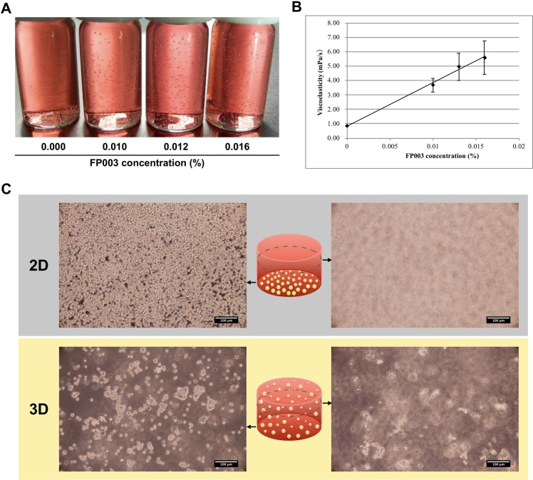
Scalable production of avian cell lines exhibits a valuable potential on therapeutic application by producing recombinant proteins and as the substrate for virus growth due to the special glycosylation occurs in avian species. Chicken primordial germ cells (cPGCs), a germinal pluripotent avian cell type, present the ability of self-renewal, an anchorage-independent cell growth and the ability to be genetically modified. This cell type could be an interesting bioreactor system for industrial purposes. This study sought to establish an expandable culture system with defined components for three-dimensional (3D) culture of cPGCs. cPGCs were cultured in medium supplemented with the functional polymer FP003. Viscoelasticity was low in this medium but cPGCs did not sediment in culture and efficiencies of space and nutrient utilization were thus enhanced and consequently their expansion was improved. The total number of cPGCs increased by 17-fold after 1 week of culture in 3D-FAot medium, an aseric defined medium containing FP003 polymer, FGF2 and Activin A as growth factors and Ovotransferrin as protein. Moreover, cPGC cell lines stably expressed the germline-specific reporter VASA:tdTOMATO, as well as other markers of cPGCs, for more than 1 month upon culture in 3D-FAot medium, indicating that the characteristics of these cells are maintained. In summary, this novel 3D culture system can be used to efficiently expand cPGCs in suspension without mechanical stirring, which is available for long-term culture and no loss of cellular properties was found. This system provides a platform for large-scale culture of cPGCs.


Conflict of interest statement

Nissan Chemical Corporation provided support in the form of salaries for authors MM, HH, and TK, but did not have any additional role in the study design, data collection and analysis, decision to publish, or preparation of the manuscript. The specific roles of these authors are articulated in the ‘author contributions’ section. This does not alter our adherence to PLOS ONE policies on sharing data and materials.

Figures

Fig 1
Fig 1. Characterization of culture medium containing FP003.
(A) Sedimentation of polystyrene beads was assessed in culture medium containing various concentrations of FP003. (B) Viscoelasticity was measured in culture medium containing different concentrations of FP003. All data are mean ± SD. (C) Culture of cPGCs in 2D and 3D media. The images show that all cPGCs settled on the bottom of the dish in 2D medium but were distributed over all surfaces in 3D medium. Scale bar: 100 μm.
Fig 2
Fig 2. Establishment of the optimal parameters for 3D culture of cPGCs in FAcs medium.
(A) Growth curves of cPGCs in 2D and 3D media over 168 hr without adding fresh medium. The 3D medium was supplemented with various concentrations of FP003. cPGCs were seeded at a density of 5 × 10⁴ cells/mL. (B) Fold increase in the total number of cPGCs grown in 2D or 3D medium for 96 hr. (C) Growth curves of cPGCs seeded at a variety of densities and cultured in medium containing 0.016% FP003 for 168 hr without adding fresh medium. The numbers of cPGCs per mL are indicated. (D) cPGCs cultured in 2D and 3D media were harvested by centrifugation at various forces. (E) cPGCs cultured in 3D medium were harvested by supplementing the culture with different amounts of citrate/PBS and then centrifuging the sample at 2000 × g. (F) Cells were harvested from 3D media. The cell pellets with different sizes in the two media were collected as indicated (arrows). All data are mean ± SEM. * p < 0.05; **** p < 0.0001.
Fig 3
Fig 3. Culture of cPGCs in serum-containing or chemically defined media.
(A) Images of cPGCs cultured in a serum-containing (FAcs) and chemically defined (FAot or FAits) media for 24, 96, and 168 hr. cPGCs were seeded at a density of 1 × 10⁴ cells/mL. Scale bar: 100 μm. (B) Fold increase in the total number of cPGCs after culture in each type of media for 24, 96, and 168 hr. Cell proliferation was assessed using the CCK-8 assay. Data are mean ± SEM, the statistical significance of difference among three groups was indicated. **** p < 0.0001. (C) Flow cytometric data. The number indicates the percentage of cells stained with an anti-SSEA-1 antibody (green). Isotype staining was performed as a control (white). (D) Percentages of SSEA-1+ cPGCs in FAcs and FAot media. Data are mean ± SEM. NS, not significant.
Fig 4
Fig 4. Expansion of cPGCs in 3D-FAcs and 3D-FAot media.
(A) Timeline of the experimental protocol. (B) Images of cPGCs cultured in 3D-FAcs and 3D-FAot media. The images were acquired by focusing on one of two surfaces, which are indicated by arrows in the cartoon. Scale bar: 100 μm. (C) Fold increase in the total number of cPGCs upon culture in 3D-FAcs and 3D-FAot media for 24, 96, and 168 hr. Data are mean ± SEM. **** p < 0.0001.
Fig 5
Fig 5. Characterization and ectopic protein expression of cPGC lines cultured for a long term in 3D media.
(A) Images showing the proliferation of vtPGCs and their expression of the germline-specific reporter tdTOMATO over 4 weeks of culture in 3D-FAcs and 3D-FAot media. Red labeling corresponds to tdTOMATO. Scale bar: 100 μm. (B) Immunofluorescence staining of SSEA-1 in vtPGCs cultured for 4 weeks in 3D-FAcs and 3D-FAot media. Green, red, and blue staining corresponds to SSEA-1, tdTOMATO, and DAPI, respectively. Scale bar: 50 μm. (C) Flow cytometric analysis of SSEA-1 and tdTOMATO expression in vtPGCs cultured for 4 weeks in 3D-FAcs and 3D-FAot media. The percentage of positively labeled vtPGCs is indicated in each graph. cPGCs were stained with mouse IgM isotype antibodies as a control. (D) RT-PCR analysis of the expression of pluripotency-related and germline-specific genes in vtPGCs cultured in 3D-FAcs and 3D-FAot media. GAPDH was used as an internal control. CEF, chicken embryonic fibroblast.
Fig 6
Fig 6. The expression of ectopic fluorescent proteins in duotonePGCs cultured in FAot or 3D-FAot medium.
(A) DuotonePGCs expressed EGFP and tdTOMATO. Scale bar: 100 μm. (B) Sedimentation of DuotonePGCs was assessed in FAot and 3D-FAot media. DuotonePGCs were largely precipitated in FAot medium and evenly distributed in 3D-FAot as the arrows indicated under the fluorescent photography. (C) Flow cytometric analysis of EGFP and tdTOMATO expression in duotonePGCs cultured in FAot and 3D-FAot media. The percentage of positively labeled cells is shown in each graph.
Fig 7
Fig 7. Graphical summary of the 3D chemically defined culture system for cPGC line by using FP003, and the comparison with the 2D condition.

References

    1. Singh H, Mok P, Balakrishnan T, Rahmat SN, Zweigerdt R. Up-scaling single cell-inoculated suspension culture of human embryonic stem cells. Stem Cell Res. 2010;4(3):165–79. 10.1016/j.scr.2010.03.001 - DOI - PubMed
    1. Amit M, Laevsky I, Miropolsky Y, Shariki K, Peri M, Itskovitz-Eldor J. Dynamic suspension culture for scalable expansion of undifferentiated human pluripotent stem cells. Nat Protoc. 2011;6(5):572–9. 10.1038/nprot.2011.325 - DOI - PubMed
    1. Otsuji TG, Bin J, Yoshimura A, Tomura M, Tateyama D, Minami I, et al. A 3D sphere culture system containing functional polymers for large-scale human pluripotent stem cell production. Stem Cell Reports. 2014;2(5):734–45. 10.1016/j.stemcr.2014.03.012 - DOI - PMC - PubMed
    1. Hiroki O, Eri N, Anna N, Hiroshi Y, Tadahiro H, Takahiro I, et al. Effective Transplantation of 2D and 3D Cultured Hepatocyte Spheroids Confirmed by Quantum Dot Imaging. Advanced Biosystems. 2018;0(0):1800137.
    1. Aihara A, Abe N, Saruhashi K, Kanaki T, Nishino T. Novel 3-D cell culture system for in vitro evaluation of anticancer drugs under anchorage-independent conditions. Cancer Sci. 2016;107(12):1858–66. 10.1111/cas.13095 - DOI - PMC - PubMed

Publication types

LinkOut - more resources