

Infection Dynamics of a Bloom-Forming Alga and Its Virus Determine Airborne Coccolith Emission from Seawater
- PMID: 30240623
- PMCID: PMC6137326
- DOI: 10.1016/j.isci.2018.07.017
Infection Dynamics of a Bloom-Forming Alga and Its Virus Determine Airborne Coccolith Emission from Seawater
Abstract
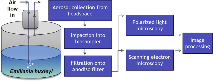
Sea spray aerosols (SSA), have a profound effect on the climate; however, the contribution of oceanic microbial activity to SSA is not fully established. We assessed aerosolization of the calcite units (coccoliths) that compose the exoskeleton of the cosmopolitan bloom-forming coccolithophore, Emiliania huxleyi. Airborne coccolith emission occurs in steady-state conditions and increases by an order of magnitude during E. huxleyi infection by E. huxleyi virus (EhV). Airborne to seawater coccolith ratio is 1:108, providing estimation of airborne concentrations from seawater concentrations. The coccoliths' unique aerodynamic structure yields a characteristic settling velocity of ∼0.01 cm s-1, ∼25 times slower than average sea salt particles, resulting in coccolith fraction enrichment in the air. The calculated enrichment was established experimentally, indicating that coccoliths may be key contributors to coarse mode SSA surface area, comparable with sea salt aerosols. This study suggests a coupling between key oceanic microbial interactions and fundamental atmospheric processes like SSA formation.
Keywords: Atmospheric Science; Biogeoscience; Earth Sciences; Marine Organism.
Copyright © 2018 The Author(s). Published by Elsevier Inc. All rights reserved.
Figures


References
-
- Aller J.Y., Kuznetsova M.R., Jahns C.J., Kemp P.F. The sea surface microlayer as a source of viral and bacterial enrichment in marine aerosols. J. Aerosol. Sci. 2005;36:801–812.
-
- Ault A.P., Moffet R.C., Baltrusaitis J., Collins D.B., Ruppel M.J., Cuadra-Rodriguez L.A., Zhao D.F., Guasco T.L., Ebben C.J., Geiger F.M. Size-dependent changes in sea spray aerosol composition and properties with different seawater conditions. Environ. Sci. Technol. 2013;47:5603–5612. - PubMed
-
- Aylward G., Findlay T. Fourth Edition. John Wiley & Sons; 1999. SI Chemical Data.
-
- Balch W.M., Holligan P.M., Ackleson S.G., Voss K.J. Biological and optical-properties of mesoscale coccolithophore blooms in the Gulf of Maine. Limnol. Oceanogr. 1991;36:629–643.
-
- Balch W.M., Kilpatrick K., Holligan P.M., Cucci T. Coccolith production and detachment by Emiliania huxleyi (Prymnesiophyceae) J. Phycol. 1993;29:566–575.
LinkOut - more resources
Full Text Sources
Other Literature Sources
Molecular Biology Databases
Research Materials
