

Muscle-specific regulation of right ventricular transcriptional responses to chronic hypoxia-induced hypertrophy by the muscle ring finger-1 (MuRF1) ubiquitin ligase in mice
- PMID: 30241514
- PMCID: PMC6150973
- DOI: 10.1186/s12881-018-0670-1
Muscle-specific regulation of right ventricular transcriptional responses to chronic hypoxia-induced hypertrophy by the muscle ring finger-1 (MuRF1) ubiquitin ligase in mice
Abstract
Background: We recently identified a role for the muscle-specific ubiquitin ligase MuRF1 in right-sided heart failure secondary to pulmonary hypertension induced by chronic hypoxia (CH). MuRF1-/- mice exposed to CH are resistant to right ventricular (RV) dysfunction whereas MuRF1 Tg + mice exhibit impaired function indicative of heart failure. The present study was undertaken to understand the underlying transcriptional alterations in the RV of MuRF1-/- and MuRF1 Tg + mice.
Methods: Microarray analysis was performed on RNA isolated from the RV of MuRF1-/-, MuRF1 Tg+, and wild-type control mice exposed to CH.
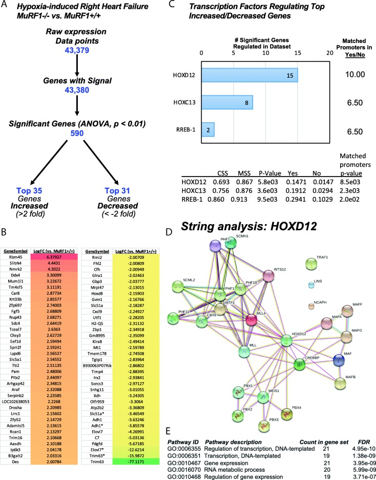
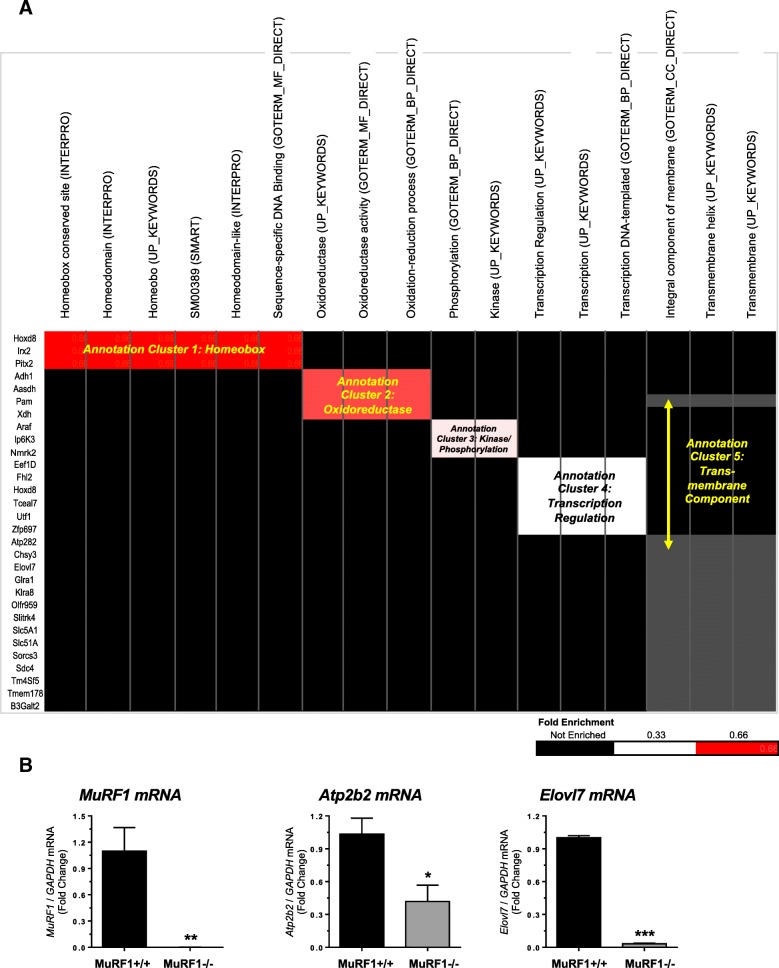
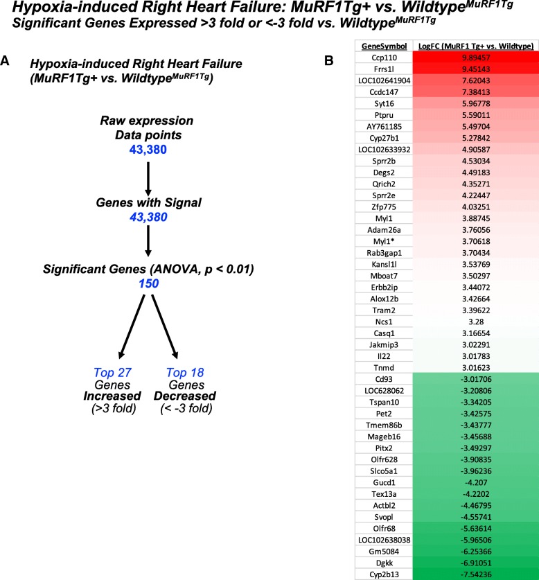
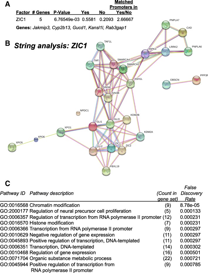
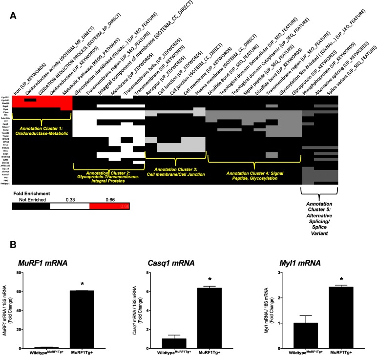
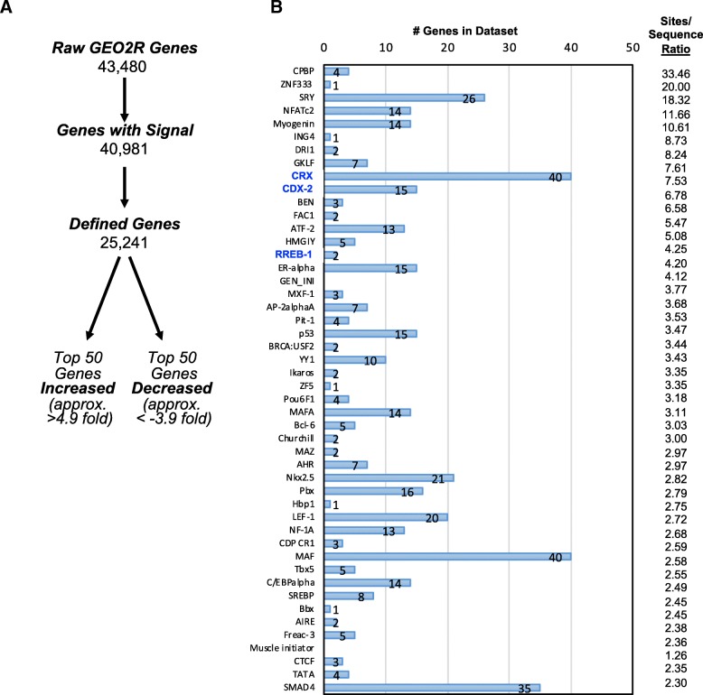
Results: MuRF1-/- RV differentially expressed 590 genes in response to CH. Analysis of the top 66 genes (> 2-fold or < - 2-fold) revealed significant associations with oxidoreductase, transcription regulation, and transmembrane component annotations. The significant genes had promoters enriched for HOXD12, HOXC13, and RREB-1 protein transcription factor binding sites. MuRF1 Tg + RV differentially expressed 150 genes in response to CH. Analysis of the top 45 genes (> 3-fold or < - 3-fold) revealed significant associations with oxidoreductase-metabolic, glycoprotein-transmembrane-integral proteins, and alternative splicing/splice variant annotations. The significant genes were enriched for promoters with ZIC1 protein transcription factor binding sites.
Conclusions: The differentially expressed genes in MuRF1-/- and MuRF1 Tg + RV after CH have common functional annotations related to oxidoreductase (including antioxidant) and transmembrane component functions. Moreover, the functionally-enhanced MuRF1-/- hearts regulate genes related to transcription, homeobox proteins, and kinases/phosphorylation. These studies also reveal potential indirect effects of MuRF1 through regulating Rreb-1, and they reveal mechanisms by which MuRF1 may transcriptionally regulate anti-oxidant systems in the face of right heart failure.
Keywords: Gene expression; Hypoxia; Microarray; MuRF1; Right heart failure.
Conflict of interest statement
Ethics approval
Mice were bred at the University of North Carolina at Chapel Hill and all the hypoxia procedures were conducted at the University of New Mexico with full approval of the UNC-Chapel Hill and University of New Mexico Institutional Animal Care and Use Committees and were carried out in compliance with the National Institutes of Health Guide for the Care and Use of Laboratory Animals.
Consent for publication
Not applicable.
Competing interests
The authors declare that they have no competing interests.
Publisher’s Note
Springer Nature remains neutral with regard to jurisdictional claims in published maps and institutional affiliations.
Figures
Similar articles
-
Muscle RING finger-1 promotes a maladaptive phenotype in chronic hypoxia-induced right ventricular remodeling.PLoS One. 2014 May 8;9(5):e97084. doi: 10.1371/journal.pone.0097084. eCollection 2014. PLoS One. 2014. PMID: 24811453 Free PMC article.
-
Muscle ring finger 1 and muscle ring finger 2 are necessary but functionally redundant during developmental cardiac growth and regulate E2F1-mediated gene expression in vivo.Cell Biochem Funct. 2014 Jan;32(1):39-50. doi: 10.1002/cbf.2969. Epub 2013 Mar 20. Cell Biochem Funct. 2014. PMID: 23512667 Free PMC article.
-
Cardiac muscle ring finger-1 increases susceptibility to heart failure in vivo.Circ Res. 2009 Jul 2;105(1):80-8. doi: 10.1161/CIRCRESAHA.109.194928. Epub 2009 Jun 4. Circ Res. 2009. PMID: 19498199 Free PMC article.
-
Skeletal muscle atrophy and the E3 ubiquitin ligases MuRF1 and MAFbx/atrogin-1.Am J Physiol Endocrinol Metab. 2014 Sep 15;307(6):E469-84. doi: 10.1152/ajpendo.00204.2014. Epub 2014 Aug 5. Am J Physiol Endocrinol Metab. 2014. PMID: 25096180 Free PMC article. Review.
-
MicroRNAs in right ventricular remodelling.Cardiovasc Res. 2017 Oct 1;113(12):1433-1440. doi: 10.1093/cvr/cvx153. Cardiovasc Res. 2017. PMID: 28957533 Review.
References
-
- Zornoff LA, Skali H, Pfeffer MA, St John Sutton M, Rouleau JL, Lamas GA, Plappert T, Rouleau JR, Moye LA, Lewis SJ, et al. Right ventricular dysfunction and risk of heart failure and mortality after myocardial infarction. J Am Coll Cardiol. 2002;39(9):1450–1455. doi: 10.1016/S0735-1097(02)01804-1. - DOI - PubMed
-
- Drake JI, Gomez-Arroyo J, Dumur CI, Kraskauskas D, Natarajan R, Bogaard HJ, Fawcett P, Voelkel NF. Chronic carvedilol treatment partially reverses the right ventricular failure transcriptional profile in experimental pulmonary hypertension. Physiol Genomics. 2013;45(12):449–461. doi: 10.1152/physiolgenomics.00166.2012. - DOI - PMC - PubMed
-
- Guihaire J, Noly PE, Schrepfer S, Mercier O. Advancing knowledge of right ventricular pathophysiology in chronic pressure overload: insights from experimental studies. Arch Cardiovasc Dis. 2015;108(10):519–29. - PubMed
-
- Ikeda S, Satoh K, Kikuchi N, Miyata S, Suzuki K, Omura J, Shimizu T, Kobayashi K, Kobayashi K, Fukumoto Y, et al. Crucial role of rho-kinase in pressure overload-induced right ventricular hypertrophy and dysfunction in mice. Arterioscler Thromb Vasc Biol. 2014;34(6):1260–1271. doi: 10.1161/ATVBAHA.114.303320. - DOI - PubMed
Publication types
MeSH terms
Substances
Grants and funding
LinkOut - more resources
Full Text Sources
Other Literature Sources
Medical
Molecular Biology Databases
Research Materials
Miscellaneous
