. 2018 Sep 21;9(1):238.
doi: 10.1186/s13287-018-0984-0.

Human chorionic villous mesenchymal stem/stromal cells protect endothelial cells from injury induced by high level of glucose

Affiliations

Human chorionic villous mesenchymal stem/stromal cells protect endothelial cells from injury induced by high level of glucose

Y S Basmaeil et al. Stem Cell Res Ther. .

Abstract

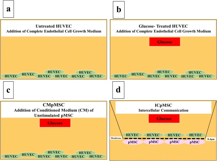
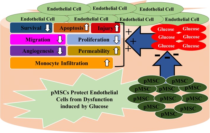
Background: Mesenchymal stem/stromal cells derived from chorionic villi of human term placentae (pMSCs) protect human endothelial cells from injury induced by hydrogen peroxide (H2O2). In diabetes, elevated levels of glucose (hyperglycaemia) induce H2O2 production, which causes the endothelial dysfunction that underlies the enhanced immune responses and adverse complications associated with diabetes, which leads to thrombosis and atherosclerosis. In this study, we examined the ability of pMSCs to protect endothelial cell functions from the negative impact of high level of glucose.

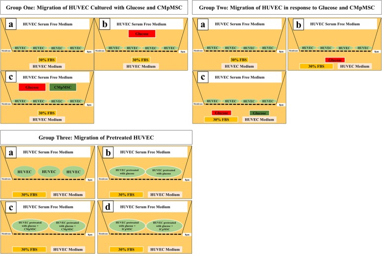
Methods: pMSCs isolated from the chorionic villi of human term placentae were cultured with endothelial cells isolated from human umbilical cord veins in the presence of glucose. Endothelial cell functions were then determined. The effect of pMSCs on gene expression in glucose-treated endothelial cells was also determined.

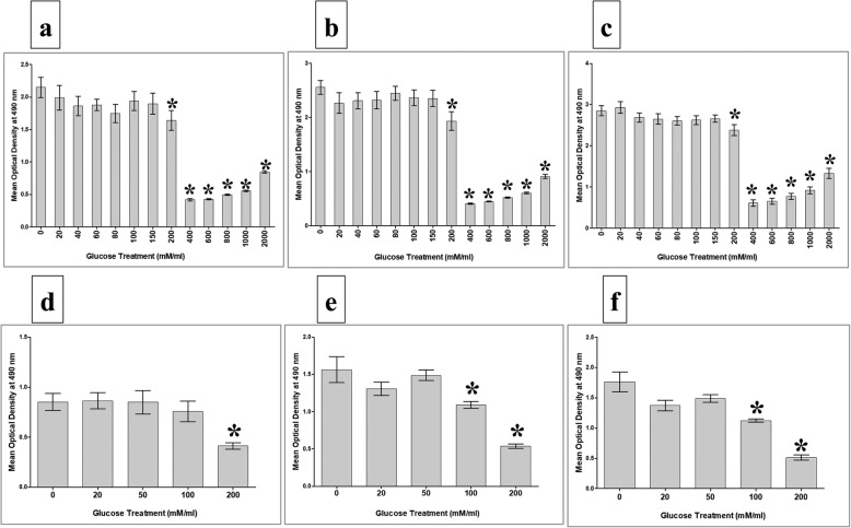
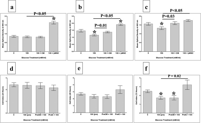
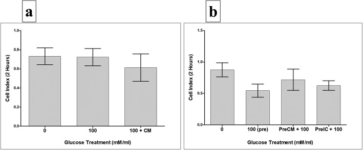
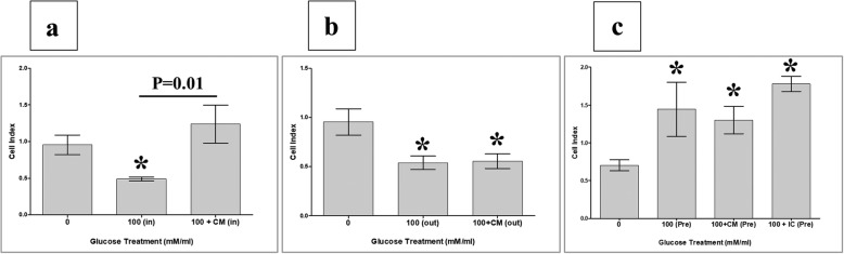
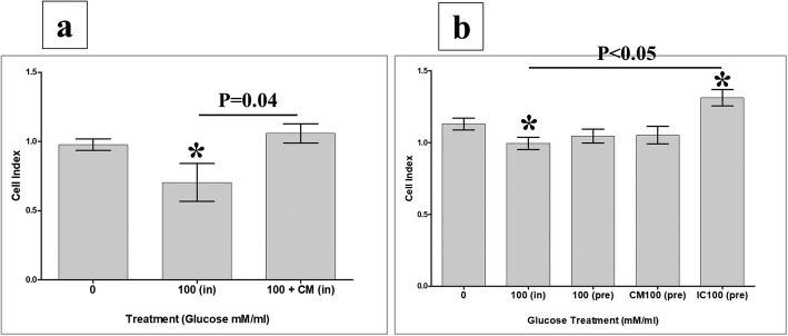
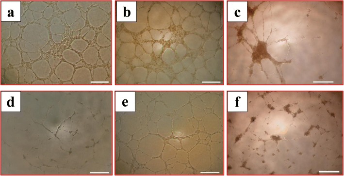
Results: pMSCs reversed the effect of glucose on key endothelial cell functions including proliferation, migration, angiogenesis, and permeability. In addition, pMSCs altered the expression of many genes that mediate important endothelial cell functions including survival, apoptosis, adhesion, permeability, and angiogenesis.

Conclusions: This is the first comprehensive study to provide evidence that pMSCs protect endothelial cells from glucose-induced damage. Therefore, pMSCs have potential therapeutic value as a stem cell-based therapy to repair glucose-induced vascular injury and prevent the adverse complications associated with diabetes and cardiovascular disease. However, further studies are necessary to reveal more detailed aspects of the mechanism of action of pMSCs on glucose-induced endothelial damage in vitro and in vivo.

Keywords: Chorionic villous mesenchymal stromal cells; Endothelial cells; Endothelium permeability; Gene expression; Glucose; Migration; Monocyte invasion; Placenta; Proliferation.


Conflict of interest statement

Ethics approval and consent to participate

The institutional review board (IRB) at King Abdulla International Medical Research Centre (KAIMRC), Saudi Arabia approved this study. Samples (i.e. placentae and umbilical cords) were obtained from uncomplicated human pregnancies (38–40 gestational weeks) following informed patient consent.

Consent for publication

Not applicable. All authors agree to publish this manuscript.

Competing interests

The authors declare that they have no competing interests.

Publisher’s Note

Springer Nature remains neutral with regard to jurisdictional claims in published maps and institutional affiliations.

Figures

Fig. 1
Fig. 1
HUVEC culture system consisted of HUVECs seeded on surface of six-well culture plate in complete HUVEC culture medium alone (untreated HUVECs) (a), or with 100 mM glucose (b), or with 100 mM glucose and 25% conditioned medium (CM) obtained from unstimulated pMSCs (c), and ICpMSC (intercellular direct contact experiment) culture system consisted of pMSCs seeded on reverse side of membrane of chamber and HUVECs seeded on upper side of membrane (d). For ICpMSC, 0.4-μm pore size transwell chamber membranes were used. In ICpMSC culture system, cells cultured at a ratio of 1 HUVEC:1 pMSC in HUVEC culture medium in presence of 100 mM glucose. In all culture systems, cells incubated for 24, 48, and 72 h at 37 °C in a cell culture incubator. CMpMSC conditioned medium of unstimulated pMSCs, HUVEC human umbilical vein endothelial cell, pMSC placental mesenchymal stem cell
Fig. 2
Fig. 2
HUVEC migration groups. Group 1: HUVECs cultured alone (a), or with 100 mM glucose (b), or with 100 mM glucose and 25% conditioned medium obtained from unstimulated pMSC culture (CMpMSC) (c) in upper chamber of CIM migration plate, while HUVEC medium with 30% FBS added to lower chamber. Group 2: HUVECs seeded in HUVEC serum-free medium in upper chamber of CIM migration plate while HUVEC medium with 30% FBS (a), or with 100 mM glucose (b), or with 100 mM glucose and 20% CMpMSC (b) added to lower chamber of migration plate. Group 3: HUVEC cultured alone (a), or with 100 mM glucose (b), or with 100 mM glucose and 25% CMpMSC (c), or with pMSCs at ratio of 1 HUVEC:1 pMSC in intercellular direct contact experiment (ICpMSC) (d). Pretreated HUVECs seeded in HUVEC serum-free medium in upper chamber of CIM migration plate while HUVEC culture medium containing 30% FBS added to lower chambers. CMpMSC conditioned medium of unstimulated pMSCs, FBS fetal bovine serum, HUVEC human umbilical vein endothelial cell, pMSC placental mesenchymal stem cell
Fig. 3
Fig. 3
Proliferation of pMSCs and HUVECs in response to various concentrations of glucose. MTS proliferation assay showed proliferation of pMSCs in response to 20, 40, 60, 80, 100, and 150 mM glucose did not significantly change as compared to glucose-untreated pMSCs after 24 h (a), 48h (b), and 72h (c) while treatment with 200–2000 mM glucose for 24 h (a), 48 h (b), and 72 h (c) significantly decreased proliferation of pMSCs as compared to glucose-untreated pMSCs. HUVEC proliferation in response to 20 and 50 mM glucose did not significantly change as compared to glucose-untreated HUVECs after 24 h (d), 48 h (e), and 72 h (f). Culture of HUVECs with 100 mM glucose for 24 h (d) did not significantly change proliferation, but significantly decreased proliferation after 48h (e) and 72 h (f) as compared to glucose-untreated pMSCs. HUVEC proliferation in response to 200 mM glucose significantly reduced as compared to glucose-untreated HUVECs after 24 h (d), 48 h (e), and 72 h (f). Each experiment performed in triplicate using pMSCs (passage 2) and HUVECs (passage 3–5) from five independent placentae and umbilical cord tissues. *p < 0.05. Bars represent standard errors
Fig. 4
Fig. 4
HUVEC proliferation in response to glucose in presence of pMSCs, or after removing glucose and pMSCs, examined after 24, 48, and 72 h in MTS assay. In response to conditioned medium (CMpMSC), pMSCs had no significant effect on HUVEC proliferation in presence of glucose after 24 h as compared to untreated or glucose-treated HUVECs (a). CMpMSC significantly increased HUVEC proliferation in presence of glucose after 48 h (b) and 72 h (c), as compared to glucose-treated but not untreated HUVECs. Cell–cell contact assay showed that, compared to glucose-untreated or treated HUVECs, pMSCs significantly increased HUVEC proliferation in presence of glucose after 24 h (a) and 48 h (b), while after 72 h (c) pMSCs significantly increased HUVEC proliferation in presence of glucose, as compared to glucose-treated but not untreated HUVECs. HUVEC proliferation after removing effects of glucose and pMSCs. HUVECs initially cultured with 100 mM glucose (100(pre)) in presence of different treatments of pMSCs (CMpMSC(PreCM + 100) and ICpMSC(PreCM + 100)) for 72 h, and then used in proliferation assay using xCELLigence real-time cell analyser. After 24 and 48 h (d, e), proliferation of HUVECs pretreated with glucose alone (100(pre)), or with CMpMSC (PreCM + 100) or ICpMSC (PreCM + 100), did not significantly change as compared to glucose-untreated HUVEC (p > 0.05). As compared to glucose-treated HUVECs (100(pre)), proliferation of HUVECs pretreated with glucose and CMpMSC (PreCM + 100), or glucose and ICpMSC (PreIC + 100), did not significantly change after 24 and 48 h (p > 0.05) (d, e). In contrast, proliferation of HUVECs pretreated with 100 mM glucose alone (100(pre)), or with CMpMSC (PreCM + 100), significantly reduced after 72 h as compared to glucose-untreated HUVEC (f). When compared with glucose-treated HUVECs (100(pre)), proliferation of HUVECs pretreated with glucose and CMpMSC (PreCM + 100) did not significantly change after 72 h of culture. In contrast, proliferation of HUVECs pretreated with glucose and ICpMSC (PreIC + 100) increased significantly after 72 h of culture as compared to glucose-treated but not untreated HUVECs (f). Each experiment performed in triplicate using HUVECs (passage 3–5) and pMSCs (passage 2) from five independent umbilical cord tissues and placentae, respectively. *P value is significant p < 0.05. Bars represent standard errors. pMSC placental mesenchymal stem cell
Fig. 5
Fig. 5
HUVEC adhesion in response to glucose and pMSCs, or after removing effects of glucose and pMSCs. HUVECs cultured with 100 mM glucose alone (100), or with 25% CMpMSC (100 + CM), and adhesion then measured using xCELLigence real-time cell analyser. After 2 h, as compared to glucose-untreated HUVECs, HUVEC adhesion in presence of glucose alone (100), or with CMpMSC (100 + CM), did not significantly change (p > 0.05) (a). Adhesion of HUVECs in presence of glucose and CMpMSC (100 + CM) did not significantly change as compared to glucose-treated HUVECs (100) after 2 h (p > 0.05) (a). HUVECs pretreated with 100 mM glucose (100(pre)) in presence of different pMSC treatments (CMpMSC (PreCM + 100) and ICpMSC (PreIC + 100)) were cultured in 16-well culture plate and adhesion measured as already indicated. After 2 h, and as compared to glucose-untreated HUVECs, preteatment of HUVECs with glucose (100(pre)), or with glucose and CMpMSC (PreCM + 100), or with glucose and ICpMSC (PreIC + 100), did not significantly change (p > 0.05) (b). Adhesion of HUVECs in presence of glucose and CMpMSC (PreCM + 100), or glucose and ICpMSC (PreIC + 100), did not significantly change as compared to glucose-treated HUVECs (100(pre)) after 2 h (p > 0.05) (b). Each experiment performed in triplicate using HUVECs (passage 3–5) and pMSCs (passage 2) from five independent umbilical cord tissues and placentae, respectively. Bars represent standard errors
Fig. 6
Fig. 6
HUVEC migration measured using xCELLigence real-time cell analyser. After 24 h, migration of HUVECs cultured with 100 mM glucose (100(in)) significantly reduced as compared to glucose-untreated HUVECs (a). As compared to glucose-untreated HUVECs, HUVEC migration in presence of glucose and CMpMSC (100 + CM(in)) did not significantly change (p > 0.05) after 24 h but migration significantly increased as compared to glucose-treated HUVECs (100(in)) (a). Migration of HUVECs in response to 100 mM glucose alone (100(out)), or with CMpMSC (100 + CM(out)) added to lower chamber of migration plate, significantly reduced as compared to glucose-untreated HUVECs after 24 h (b). As compared to glucose-treated HUVECs (100(out)), HUVEC migration in response to glucose and CMpMSC (100 + CM(out)) did not significantly change after 24 h as compared to glucose-treated HUVECs (100(out)) (b). After 24 h, migration of HUVECs pretreated with glucose alone (100(pre), or with glucose and CMpMSC (100 + CM(Pre)), or with glucose and ICpMSC (100 + IC(Pre)), significantly increased as compared to glucose-treated HUVECs (100(pre)) (c). After 24 h, as compared to glucose-treated HUVECs (100(pre)), migration of HUVECs pretreated with 100 mM glucose and CMpMSC (100 + CM(Pre)), or with 100 mM glucose and ICpMSC (100 + IC(Pre)), did not significantly change (p > 0.05) (c). Each experiment performed in triplicate using HUVECs (passage 3–5) and pMSCs (passage 2) from five independent umbilical cord tissues and placentae, respectively. *P value is significant < 0.05. Bars represent standard errors
Fig. 7
Fig. 7
HUVEC permeability under effects of glucose and pMSCs examined by adding monocytes to monolayer of HUVECs and assessing invasion of monocytes through HUVEC monolayer by xCELLigence real-time system. Increased invasion defined as reduction in cell index due to infiltration of HUVEC monolayer by monocytes, causing detachment of HUVECs, while increased cell index defines reduction in cell invasion. In presence of 100 mM/ml glucose (100(in)), monocyte invasion of HUVEC monolayer significantly increased after 10 h as compared to glucose-untreated HUVECs (a). After 10 h and as compared to glucose-untreated HUVECs, monocyte invasion in presence of glucose and CMpMSC (100 + CM(in)) significantly reduced but not significantly changed as compared to glucose-untreated HUVECs (a). Monocyte invasion through monolayer of HUVECs pretreated with glucose alone (100(pre)), or with glucose and CMpMSC (100 + CM(pre)), not significantly changed after 10 h as compared to glucose-untreated HUVECs, while addition of ICpMSC (100 + IC(pre)) significantly reduced monocyte invasion as compared to glucose-treated (100(pre)) or untreated HUVECs (b). Each experiment performed in triplicate using HUVECs (passage 3–5) and pMSCs (passage 2) from five independent umbilical cord tissues and placentae, respectively. *P value is significant < 0.05. Bars represent standard errors
Fig. 8
Fig. 8
HUVEC tubule formation in presence of glucose and pMSCs. After 14 h, glucose-untreated pMSCs (a) and HUVECs cultured with 25% CMpMSC (b) able to form tube networks. HUVECs cultured with pMSCs alone did not form extensive tubule networks (c), and with 100 mM glucose alone (d) were unable to form tube networks. HUVECs cultured with 100 mM glucose and 25% CMpMSC (e) able to form tube networks, while culturing HUVECs with 100 mM glucose and pMSCs (f) failed to form extensive tube networks. Each experiment performed in triplicate using HUVECs (passage 3–5) and pMSCs (passage 2) from five independent umbilical cord tissues and placentae, respectively
Fig. 9
Fig. 9
Proposed effects of placental mesenchymal stem cells (pMSCs) on modifying negative impact of glucose on endothelial cell functions. pMSCs prevent endothelial cell injury and apoptosis from glucose and reverse inhibitory effects of glucose on endothelial cell survival, proliferation, migration, and angiogenesis. pMSCs also reduce stimulatory effects of glucose on endothelial cell permeability and monocyte infiltration through endothelial cells

Similar articles

Cited by

References

    1. Prentki M, Nolan CJ. Islet beta cell failure in type 2 diabetes. J Clin Invest. 2006;116(7):1802–1812. doi: 10.1172/JCI29103. - DOI - PMC - PubMed
    1. Beckman JA, Creager MA, Libby P. Diabetes and atherosclerosis: epidemiology, pathophysiology, and management. JAMA. 2002;287(19):2570–2581. doi: 10.1001/jama.287.19.2570. - DOI - PubMed
    1. Morel O, Kessler L, Ohlmann P, Bareiss P. Diabetes and the platelet: toward new therapeutic paradigms for diabetic atherothrombosis. Atherosclerosis. 2010;212(2):367–376. doi: 10.1016/j.atherosclerosis.2010.03.019. - DOI - PubMed
    1. Ferreiro JL, Angiolillo DJ. Diabetes and antiplatelet therapy in acute coronary syndrome. Circulation. 2011;123(7):798–813. doi: 10.1161/CIRCULATIONAHA.109.913376. - DOI - PubMed
    1. Paneni F, Beckman JA, Creager MA, Cosentino F. Diabetes and vascular disease: pathophysiology, clinical consequences, and medical therapy: part I. Eur Heart J. 2013;34(31):2436–2443. doi: 10.1093/eurheartj/eht149. - DOI - PMC - PubMed

Publication types

MeSH terms

LinkOut - more resources