

Associations of cardiovascular fat radiodensity and vascular calcification in midlife women: The SWAN cardiovascular fat ancillary study
- PMID: 30241697
- PMCID: PMC6295258
- DOI: 10.1016/j.atherosclerosis.2018.09.001
Associations of cardiovascular fat radiodensity and vascular calcification in midlife women: The SWAN cardiovascular fat ancillary study
Abstract
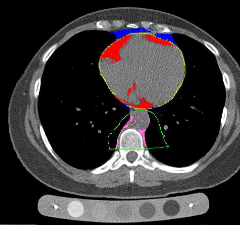
Background and aims: Fat radiodensity, measured via CT Hounsfield units (HU), is a potential marker of fat quality. We sought to determine the cross-sectional associations of total heart fat (TAT) and aortic perivascular fat (PVAT) radiodensity with cardiovascular risk factors, coronary artery calcification (CAC), and aortic calcification (AC) in midlife women.
Methods: Fat radiodensity, CAC, and AC were quantified using CT scans. A total of 528 women (mean age: 50.9 ± 2.9 years; 37% Black) were included in analyses.
Results: Women in the lowest TAT radiodensity tertile were more likely to have adverse cardiovascular risk factors. Independent of cardiovascular risk factors, women in the middle and high TAT radiodensity tertiles were less likely to have CAC (OR (95% CI): 0.32 (0.18, 0.59); 0.43 (0.24, 0.78), respectively) compared with women in the lowest TAT radiodensity tertile. Although adjusting for BMI attenuated the overall association, women in the middle TAT radiodensity tertile remained at significantly lower odds of CAC when compared to the low radiodensity tertile, 0.47 (0.24, 0.93), p=0.03. No significant associations were found for PVAT radiodensity and calcification measures in multivariable analysis.
Conclusions: Lower TAT radiodensity was associated with a less favorable cardiometabolic profile. Women with mid-range TAT radiodensity values had a lower odds of CAC presence, independent of CVD risk factors and BMI. More research is necessary to understand radiodensity as a surrogate marker of fat quality in midlife women.
Keywords: Cardiovascular; Fat radiodensity; Midlife women; Vascular calcification.
Copyright © 2018 Elsevier B.V. All rights reserved.
Conflict of interest statement
Conflicts of Interest
Dr. Hanley has nothing to disclose; Dr. Shields has nothing to disclose, Dr. El Khoudary reports grants from AHA, during the conduct of the study; Dr. Matthews has nothing to disclose; Dr. Brooks reports grants from Gilead Science Inc, outside the submitted work; Dr. Janssen reports grants from NIH, during the conduct of the study; Dr. Budoff reports grants from NIH, during the conduct of the study; grants from GE, outside the submitted work; Dr. Sekikawa has nothing to disclose; Dr. Mulukutla has nothing to disclose.
Figures
Comment in
-
Thoracic adipose tissue density as a novel marker of increased cardiovascular risk.Atherosclerosis. 2018 Dec;279:91-92. doi: 10.1016/j.atherosclerosis.2018.10.002. Epub 2018 Oct 6. Atherosclerosis. 2018. PMID: 30327129 No abstract available.
References
-
- Kershaw EE and Flier JS, Adipose tissue as an endocrine organ, The Journal of clinical endocrinology and metabolism, 2004;89:2548–2556. - PubMed
-
- Yudkin JS, Eringa E and Stehouwer CD, “Vasocrine” signalling from perivascular fat: a mechanism linking insulin resistance to vascular disease, Lancet, 2005;365:1817–1820. - PubMed
-
- Iacobellis G and Barbaro G, The double role of epicardial adipose tissue as pro- and anti-inflammatory organ, Horm Metab Res, 2008;40:442–445. - PubMed
-
- Ahmadi N, Nabavi V, Yang E, Hajsadeghi F, Lakis M, et al., Increased epicardial, pericardial, and subcutaneous adipose tissue is associated with the presence and severity of coronary artery calcium, Acad Radiol, 2010;17:1518–1524. - PubMed
-
- Bettencourt N, Toschke AM, Leite D, Rocha J, Carvalho M, et al., Epicardial adipose tissue is an independent predictor of coronary atherosclerotic burden, Int J Cardiol, 2012;158:26–32. - PubMed
Publication types
MeSH terms
Grants and funding
LinkOut - more resources
Full Text Sources
Other Literature Sources
Medical
