Clinical Trial
. 2019 Jan 1;25(1):110-124.
doi: 10.1158/1078-0432.CCR-18-1763. Epub 2018 Sep 21.

Immunotherapy Targeting HPV16/18 Generates Potent Immune Responses in HPV-Associated Head and Neck Cancer

Affiliations
Clinical Trial

Immunotherapy Targeting HPV16/18 Generates Potent Immune Responses in HPV-Associated Head and Neck Cancer

Charu Aggarwal et al. Clin Cancer Res. .

Abstract

Purpose: Clinical responses with programmed death (PD-1) receptor-directed antibodies occur in about 20% of patients with advanced head and neck squamous cell cancer (HNSCCa). Viral neoantigens, such as the E6/E7 proteins of HPV16/18, are attractive targets for therapeutic immunization and offer an immune activation strategy that may be complementary to PD-1 inhibition.

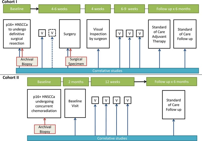
Patients and methods: We report phase Ib/II safety, tolerability, and immunogenicity results of immunotherapy with MEDI0457 (DNA immunotherapy targeting HPV16/18 E6/E7 with IL12 encoding plasmids) delivered by electroporation with CELLECTRA constant current device. Twenty-two patients with locally advanced, p16+ HNSCCa received MEDI0457.

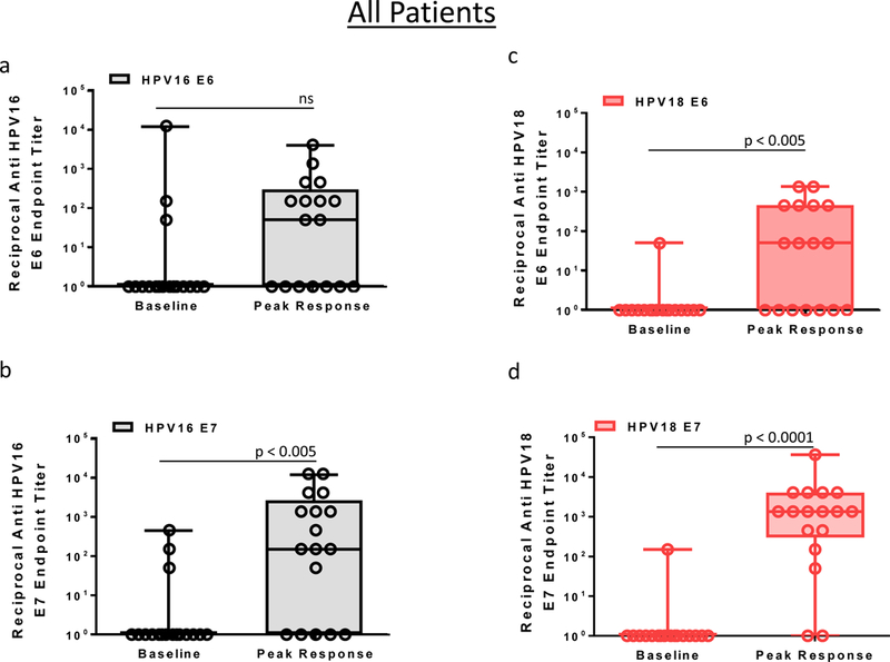
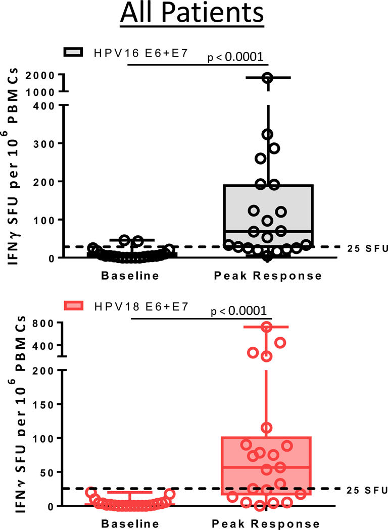
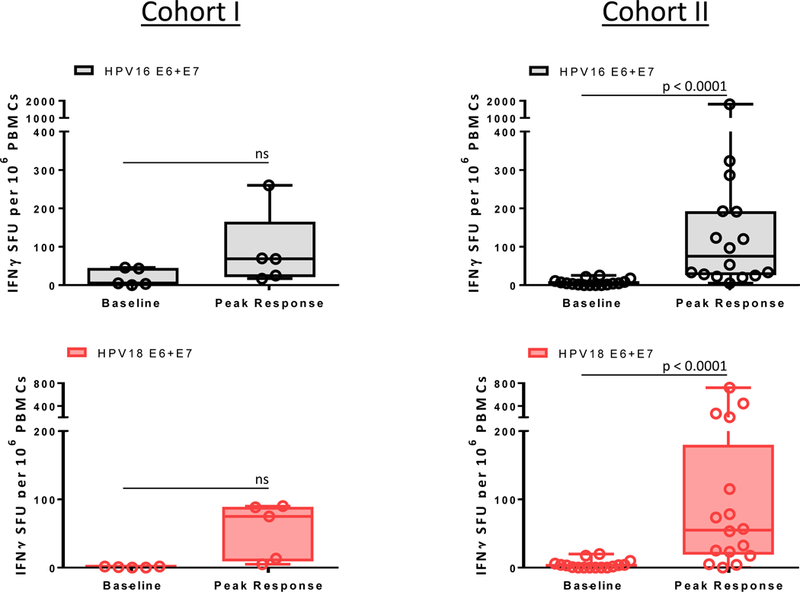
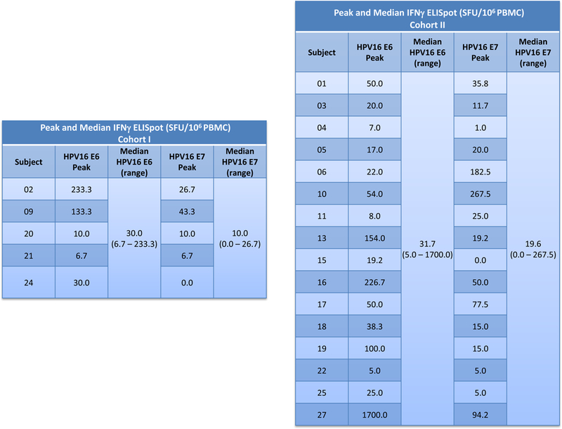
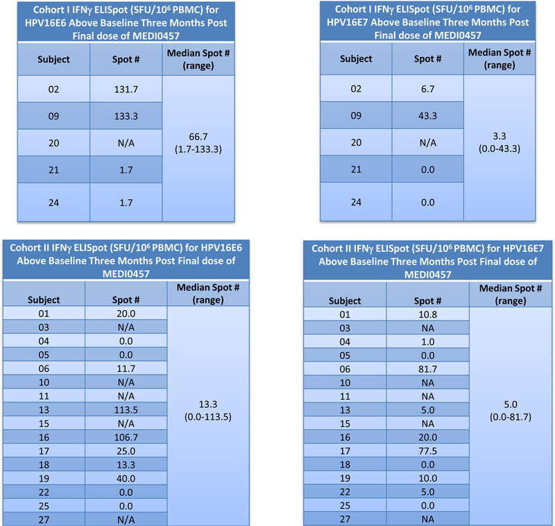
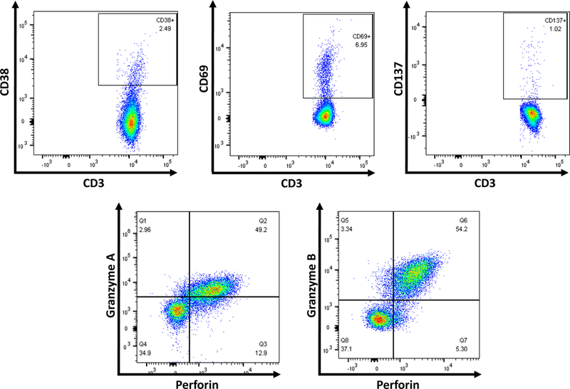
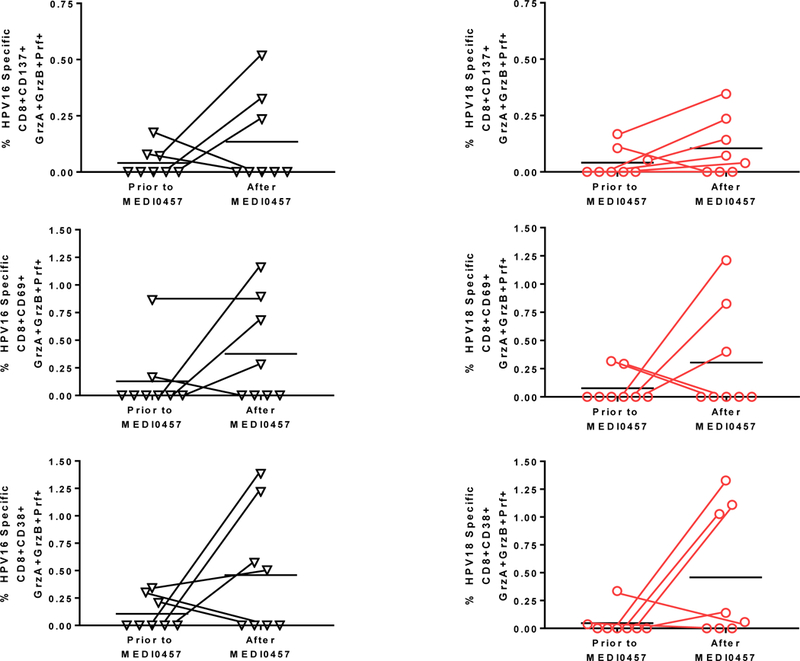
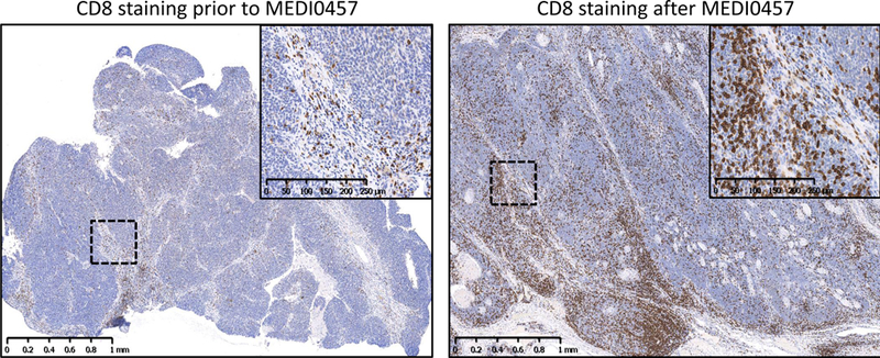
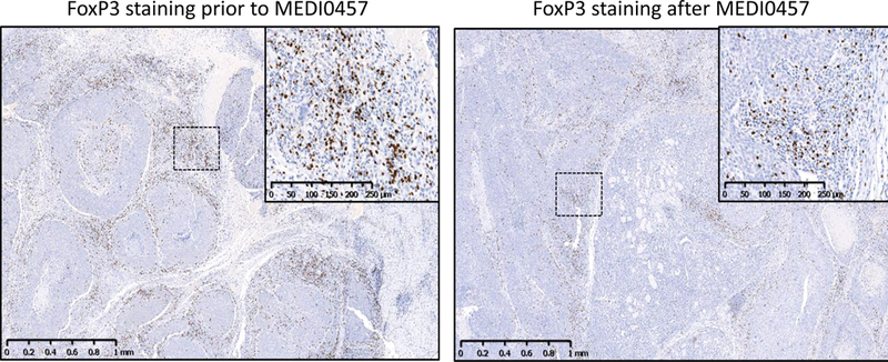
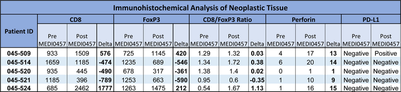
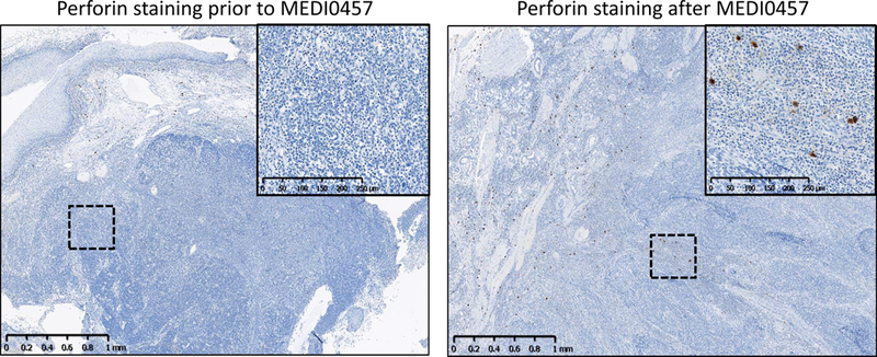
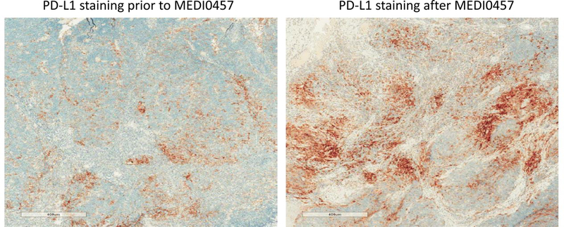
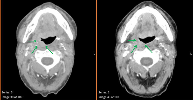
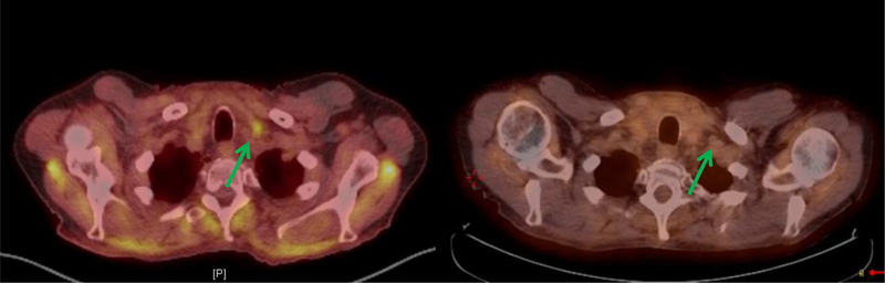
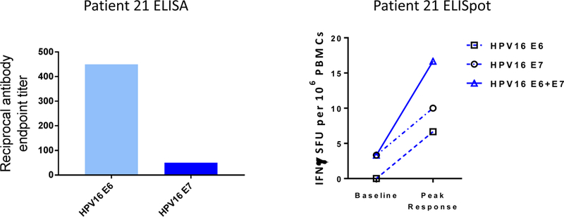
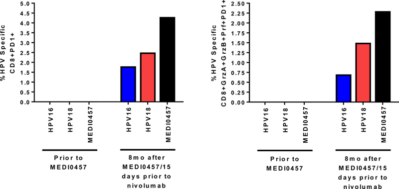
Results: MEDI0457 was associated with mild injection site reactions, but no treatment-related grade 3-5 adverse events (AE) were noted. Eighteen of 21 evaluable patients showed elevated antigen-specific T-cell activity by IFNγ ELISpot, and persistent cellular responses surpassing 100 spot-forming units (SFUs)/106 peripheral blood mononuclear cells (PBMCs) were noted out to 1 year. Induction of HPV-specific CD8+ T cells was observed. MEDI0457 shifted the CD8+/FoxP3+ ratio in 4 of 5 post immunotherapy tumor samples and increased the number of perforin+ immune infiltrates in all 5 patients. One patient developed metastatic disease and was treated with anti-PD-1 therapy with a rapid and durable complete response. Flow-cytometric analyses revealed induction of HPV16-specific PD-1+ CD8+ T cells that were not found prior to MEDI0547 (0% vs. 1.8%).

Conclusions: These data demonstrate that MEDI0457 can generate durable HPV16/18 antigen-specific peripheral and tumor immune responses. This approach may be used as a complementary strategy to PD-1/PD-L1 inhibition in HPV-associated HNSCCa to improve therapeutic outcomes.


Conflict of interest statement

Conflict of Interest Disclosures:Charu Aggarwal reported consulting or advisory roles with Genentech, Bristol-Myers Squibb, Lilly, and Celgene; and institutional research funding from Genetech/Roche, Incyte, Macrogenics, and Merck Sharp & Dohme. Roger B. Cohen reported honoraria from Bristol-Myers Squibb; a consulting or advisory role with Heat Biologics, Takeda, Cerulean Pharma, Kolltan Pharmacueticals, Zymeworks, and Pfizer; institutional research funding from Heat Biologics, Macrogenetics, Merck, Takeda, Cleave Biosciences, and Celldex; and travel, accommodations, or expenses from Heat Biologics, Takeda, Kolltan Pharmaceuticals, Cerulean Pharma, Zymeworks, Bristol-Meyers Squibb, and Pfizer. J.M. Bauml reported consulting or advisory roles with Clovis Oncology, Bristol-Myers Squibb, Merck, AstraZeneca, Genentech, Celgene, Boehringer Ingelheim, and Guardant Health; and institutional research funding from Merck, Carevive Systems, Novartis, Incyte, Bayer, and Janssen. GS Weinstein and Alexander Lin have no relevant disclosures. Russell Vang has no relevant disclosures. David B. Weiner received a SRA research grant from Inovio Pharmaceuticals, has received speakers honoraria from Inovio Pharmaceuticals, GeneOne, Astrazeneca, BMGF, he has ownership interest (including patents) in Inovio Pharmaceuticals and is a consultant/advisory board member for Inovio Pharmaceuticals, a consultant for GeneOne and Astrazeneca and Merck. All authors whose listed association is Inovio Pharmaceuticals (MPM, KAK, AJS, DMK, JB, LS, ST, AA, KD, DM, MD, SO, SD, IC, MLB) and Medimmune (ME, RK) are employees of that entity, own stock or stock options relative to that entity and may own one or more patents relating to the drugs being described in this publication.

Figures

Figure 1:
Figure 1:
Trial Schema. Patients were enrolled in 2 cohorts: Cohort I: patients that underwent definitive surgical resection. Patients could receive 1–2 doses of MEDI0457 prior to surgery. About 4 weeks post-surgery, if adequate healing was documented by the surgeon, the patient resumed immunotherapy dosing every 3 weeks, for a total of 4 doses. Patients then went on to receive standard adjuvant therapy based on pathologic features. Cohort II: patients started immunotherapy 2 months after completion of chemoradiation. Blue arrows denote the timing of peripheral immune analyses. Red arrows denote the timing of tissue analyses. V= visits for immunotherapy administration.
Fig 2A:
Fig 2A:
Treatment with MEDI0457 induces the generation of HPV16 and HPV18 specific antibodies. Immunoglobulin G (IgG) responses at baseline, and peak response post immunotherapy with MEDI0457 (a to d) HPV16 E6 (a), HPV16 E7 (b), HPV18 E6 (c), and HPV18 E7 (d) measured by ELISA. Peak response is defined as the highest titer noted against the graphed antigen at any time point post treatment. Statistical significance is noted where confirmed for a given comparison.
Fig 2B:
Fig 2B:
IFN-γ ELISpot responses to HPV16 and HPV18 antigens are elevated after treatment with MEDI0457 when assessed irrespective of cohort. Number of antigen-specific IFN-γ–secreting cells in response to stimulation with HPV16 E6 and E7 pools (top panel) and HPV18 E6 and E7 pools (lower panel) at baseline and peak response measured by IFN-γ ELISpot assay for all patients in the trial irrespective of cohort. The dashed line represents a cutoff below which responses are considered “low”. Peak response is defined as the highest SFU reading noted against the graphed antigen at any time point post treatment. Statistical significance is noted where confirmed for a given comparison.
Figure 2C:
Figure 2C:
IFN-γ ELISpot responses to HPV16 and HPV18 antigens are elevated after treatment with MEDI0457 when assessed by cohort. Number of antigen-specific IFN-γ–secreting cells in response to stimulation with HPV16 E6 and E7 pools in cohort I (upper left) and Cohort II (upper right) or HPV18 E6 and E7 pools in Cohort 1 (lower left) and Cohort II (lower right). Peak response is defined as the highest SFU reading noted against the graphed antigen at any time point post treatment. Statistical significance is noted where confirmed for a given comparison.
Figure 2D:
Figure 2D:
Peak per-subject and median (plus range) IFN-γ ELISpot responses to HPV16 E6 and E7 antigens after treatment with MEDI0457. For Cohort I (left panel) and Cohort II (right panel) at the “peak” timepoint post treatment.
Figure 2E:
Figure 2E:
Persistent per subject and median (plus range) IFN-γ ELISpot responses to HPV16 E6 and E7 antigens after treatment with MEDI0457. For Cohort I (top panels) and Cohort II (bottom panels) the time is three months post treatment. Where sample was unable to be recovered for testing “N/A” is listed.
Fig 3A:
Fig 3A:
MEDI0457 induces HPV16 and HPV18 CD8+ T cells with phenotypic lytic markers as gauged by flow cytometry. A: Flow cytometric assessments of CD8+ T cell compartment for cell surface markers CD38, CD69, and CD137 and lytic markers granzyme A (GrzA), granzyme B (GrzB) and perforin (Prf). Representative gates are shown.
Figure 3B:
Figure 3B:
Induction of antigen specific CD8+ T cell populations expressing GrzA, GrzB and Prf; HPV 16 specific (left column, black triangles) and HPV 18 specific (right column, red circles). Flow cytometry was performed on patients who had sufficient recoverable PBMCs (Total n = 8, n=2 from Cohort I, n=6 from Cohort 2). Each triangle or circle represents an individual patient. The line that connects triangles or circles shows the increase or decrease from the timepoint prior to dosing with MEDI0457 to the timepoint following dosing with MEDI0457. Although mean frequencies increase for all comparisons, the increases are not statistically significant.
Fig 4(A):
Fig 4(A):
Treatment with MEDI0457 modulates immune infiltration into tumor tissues. Immunohistochemical analysis of neoplastic tissue in paired samples pre- and post-MEDI0457,CD8
Fig 4B:
Fig 4B:
Treatment with MEDI0457 modulates immune infiltration into tumor tissues. Immunohistochemical analysis of neoplastic tissue in paired samples pre- and post-MEDI0457,FoxP3.
Figure 4C:
Figure 4C:
Numbers for cells staining positive for CD8, FoxP3, Perforin and PD-L1 per mm2 neoplastic tissue are listed for each patient along with CD8/FoxP3 ratio.
Fig 4D:
Fig 4D:
Immunohistochemical analysis of neoplastic tissue in paired samples pre- and post-MEDI0457 for Perforin
Fig 4E:
Fig 4E:
PD-L1; PD-L1 status of positive is assigned to cases with total percent of tumor cells with membrane staining of any intensity of greater than or equal to 25%
Fig 5(A):
Fig 5(A):
Treatment of a patient with progressive disease with dermal and lymph node metastases (patient 21) with nivolumab resulted in radiographic complete response CT neck with IV contrast
Fig 5(B):
Fig 5(B):
PET CT scan images pre- and 6 weeks post- nivolumab. There is abnormal tissue in the right oropharynx involving the tonsil, lateral oropharyngeal wall, right glossotonsillar sulcus (green arrows) and right base of tongue extending to the midline. At 6 weeks post nivolumab, there was interval reduction in this solid enhancing tissue component. PET images showed interval resolution of a hypermetabolic left supraclavicular lymph node (green arrow) seen on the prior exam.
Fig 5C:
Fig 5C:
Assessment of peripheral immune responses in patient 21; ELISA antibody responses (left panel) and cellular responses by IFNGamma ELISpot (right panel).
Fig 5D:
Fig 5D:
Analysis of CD8+T cells specific for HPV16 E6 and E7 peptides, HPV18 E6 and E7 peptides or both (MEDI0457) prior to and post dosing with MEDI0457. Frequencies of PD-1+ (left panel) and GrzA+GrzB+Prf+PD-1+ (right panel) of all peripheral CD8+ T cells specific for HPV 16 peptides (blue bars), HPV 18 peptides (red bars) or pooled HPV16 and HPV18 peptides (MEDI0457, black bars).

Similar articles

Cited by

References

    1. Siegel RL, Miller KD, Jemal A. Cancer Statistics, 2017. CA: a cancer journal for clinicians. 2017;67(1):7–30. Epub 2017/01/06. - PubMed
    1. Varilla V, Atienza J, Dasanu CA. Immune alterations and immunotherapy prospects in head and neck cancer. Expert opinion on biological therapy. 2013;13(9):1241–56. Epub 2013/06/25. - PubMed
    1. Sun W, Li WJ, Wu CY, Zhong H, Wen WP. CD45RA-Foxp3high but not CD45RA+Foxp3low suppressive T regulatory cells increased in the peripheral circulation of patients with head and neck squamous cell carcinoma and correlated with tumor progression. Journal of experimental & clinical cancer research : CR. 2014;33:35. Epub 2014/04/26. - PMC - PubMed
    1. Mandal R, Senbabaoglu Y, Desrichard A, Havel JJ, Dalin MG, Riaz N, et al. The head and neck cancer immune landscape and its immunotherapeutic implications. JCI insight. 2016;1(17):e89829. Epub 2016/10/26. - PMC - PubMed
    1. Chaturvedi AK, Engels EA, Pfeiffer RM, Hernandez BY, Xiao W, Kim E, et al. Human papillomavirus and rising oropharyngeal cancer incidence in the United States. Journal of clinical oncology : official journal of the American Society of Clinical Oncology. 2011;29(32):4294–301. Epub 2011/10/05. - PMC - PubMed

Publication types

MeSH terms