. 2018 Oct 31;38(5):BSR20181043.
doi: 10.1042/BSR20181043. Print 2018 Oct 31.

Bakuchiol protects against pathological cardiac hypertrophy by blocking NF-κB signaling pathway

Affiliations

Bakuchiol protects against pathological cardiac hypertrophy by blocking NF-κB signaling pathway

Zheng Wang et al. Biosci Rep. .

Abstract

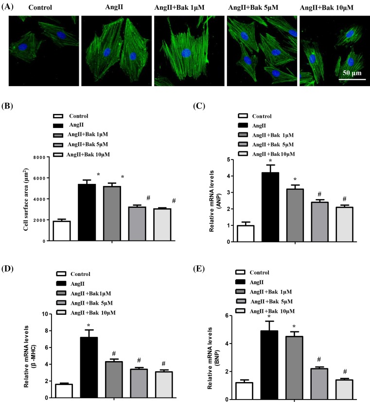
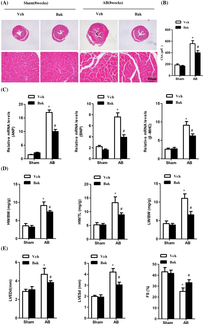
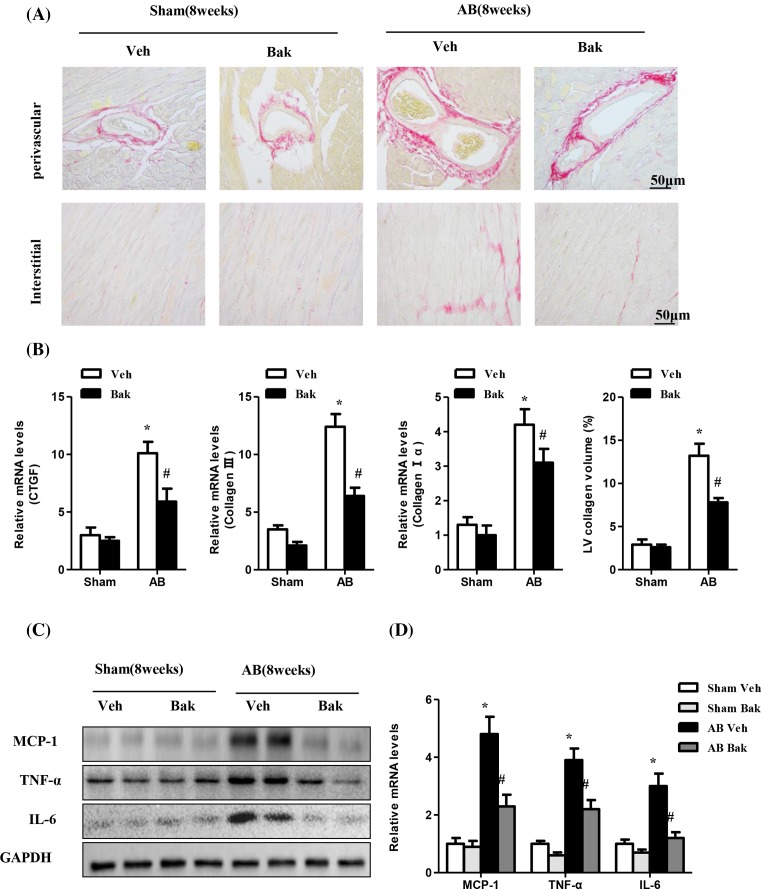
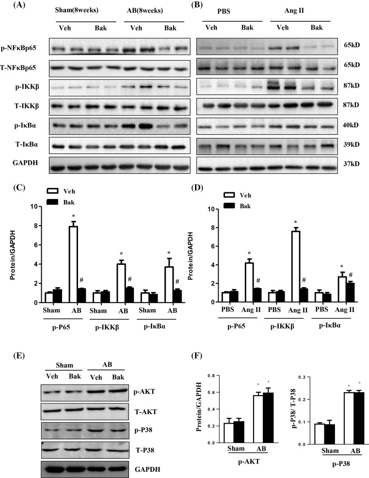
Bakuchiol (Bak), a monoterpene phenol isolated from the seeds of Psoralea corylifolia, has been widely used to treat a large variety of diseases in both Indian and Chinese folkloric medicine. However, the effects of Bak on cardiac hypertrophy remain unclear. Therefore, the present study was designed to determine whether Bak could alleviate cardiac hypertrophy. Mice were subjected to aortic banding (AB) to induce cardiac hypertrophy model. Bak of 1 ml/100 g body weight was given by oral gavage once a day from 1 to 8 weeks after surgery. Our data demonstrated for the first time that Bak could attenuate pressure overload-induced cardiac hypertrophy and could attenuate fibrosis and the inflammatory response induced by AB. The results further revealed that the effect of Bak on cardiac hypertrophy was mediated by blocking the activation of the NF-κB signaling pathway. In vitro studies performed in neonatal rat cardiomyocytes further proved that the protective effect of Bak on cardiac hypertrophy is largely dependent on the NF-κB pathway. Based on our results, Bak shows profound potential for its application in the treatment of pathological cardiac hypertrophy, and we believe that Bak may be a promising therapeutic candidate to treat cardiac hypertrophy and heart failure.

Keywords: Bakuchiol; NF kappa B; aortic banding; cardiac hypertrophy; cardiomyocytes.


Conflict of interest statement

The authors declare that there are no competing interests associated with the manuscript.

Figures

Figure 1
Figure 1. Bak inhibits cardiomyocyte hypertrophy in vitro
(A) Representative images of NRCMs pretreated with different concentrations of Bak for 48 h and subsequently treated with 1 μM Ang II for 24 h (n = 3 independent experiments; blue, nucleus; green, α-actinin; scale bar, 50 μm). (B) Individual cell surface area of at least 100 NRCMs per group were traced and compared between the indicated groups (*P<0.05 versus control; #P<0.05 versus Ang II-treated cells). (CE) The relative mRNA levels of the hypertrophic markers ANP, BNP, and β-MHC were analyzed by real-time PCR and compared between the indicated groups (*P<0.05 versus control; #P<0.05 versus Ang II-treated cells).
Figure 2
Figure 2. Bak attenuates pressure overload-induced cardiac hypertrophy and improves impaired cardiac function
(A) Representative images of sections subjected to HE staining from vehicle (Veh) or Bak-treated mice 8 weeks after sham or AB surgery (n = 5–6 mice per experimental group; scale bar, 50 μm). (B) The statistical results of cardiomyocyte CSAs in each group (n≥100 cells; *P<0.05 versus vehicle/sham, #P<0.05 versus vehicle/AB after AB). (C) Real-time PCR analyses of the hypertrophy markers ANP, BNP, and β-MHC induced by AB or sham surgery in each group of mice (n=5; *P<0.05 versus vehicle/sham; #P<0.05 versus vehicle/AB after AB). (D) The statistical results of the HW/BW, HW/TL, and LW/BW ratios in the indicated groups (n=5; *P<0.05 versus vehicle/sham; #P<0.05 versus vehicle/AB after AB). (E) Statistical results of the echocardiographic parameters of the four groups of mice after 8 weeks of AB or sham operations (n=5; *P<0.05 versus vehicle/sham; #P<0.05 versus vehicle/AB after AB).
Figure 3
Figure 3. Bak attenuates fibrosis and inflammatory responses induced by AB
(A) Representative images of sections subjected to PSR staining from Veh- or Bak-treated mice 8 weeks after sham or AB surgery (n = 5–6 mice per experimental group; scale bar, 50 μm). (B) The statistical results of LV collagen volume in each group (n≥25 fields from 5 mice per experimental group; *P<0.05 versus vehicle/sham; #P<0.05 versus vehicle/AB after AB). (C) Representative Western blots and quantitative results (D) showing MCP-1, TNFα, and IL-6 expression in heart tissues of mice in the indicated groups (n=5; *P<0.05 versus vehicle/sham; #P<0.05 versus vehicle/AB after AB).
Figure 4
Figure 4. Bak mediates cardiac hypertrophy through the inhibition of the NF-κB pathway
(A) Representative Western blotting analysis and quantitative results (C) showing phosphorylated and total NF-κB p65, IKKβ, and inhibitor of NF-κB a (IκBa) expression in heart tissues of mice in the indicated groups (n=5; *P<0.05 versus vehicle/sham; #P<0.05 versus vehicle/AB after AB). (B) Representative Western blotting analysis and quantitative results (D) showing phosphorylated and total NF-κB p65, IKKβ and IκBa expression in NRCMs treated with Bak (10 μM) for 48 h subsequently treated with 1 μM Ang II for 24 h. (*P<0.05 versus control; #P<0.05 versus AngII-treated cells) (E and F). Representative Western blotting analysis (E) and quantitative results (F) showing phosphorylated and total Akt and P38 expression in heart tissues of mice in the indicated groups (n=5; *P<0.05 versus vehicle/sham; #P<0.05 versus vehicle/AB after AB).
Figure 5
Figure 5. The protective effect of Bak on cardiac hypertrophy is largely dependent on the NF-κB pathway
(A) Representative Western blotting analysis and quantitative results showing phosphorylated NF-κB p65 (*P<0.05 versus control). (B) Representative images of NRCMs that were pretreated with PDTC for 1 h and those that were subsequently treated with 1 μM Ang II for 48 h after being treated with Bak for 24 h. (C) Quantitation of the cell surface area (n=100 cells). (D) Real-time PCR analysis of the hypertrophy markers ANP, BNP, and β-MHC (*P<0.05 versus control; #P<0.05 versus Ang II-treated cells).

References

    1. Frey N. and Olson E.N. (2003) Cardiac hypertrophy: the good, the bad and the ugly. Annu. Rev. Physiol. 65, 45–79 10.1146/annurev.physiol.65.092101.142243 - DOI - PubMed
    1. Bernardo B.C., Weeks K.L., Pretorius L. and McMullen J.R. (2010) Molecular distinction between physiological and pathological cardiac hypertrophy: Experimental findings and therapeutic strategies. Pharmacol. Ther. 128, 191–227 10.1016/j.pharmthera.2010.04.005 - DOI - PubMed
    1. Ruwhof C. and Van Der Laarse A. (2000) Mechanical stress-induced cardiac hypertrophy: mechanisms and signal transduction pathways. Cardiovasc. Res. 47, 23–37 10.1016/S0008-6363(00)00076-6 - DOI - PubMed
    1. Hunter J.J. and Chien K.R. (1999) Mechanisms of disease - signaling pathways for cardiac hypertrophy and failure. N. Engl. J. Med. 341, 1276–1283 10.1056/NEJM199910213411706 - DOI - PubMed
    1. Amin R., Muthuramu I., Aboumsallem J.P., Mishra M., Jacobs F. and De Geest B. (2017) Selective HDL-Raising human Apo A-I gene therapy counteracts cardiac hypertrophy, reduces myocardial fibrosis, and improves cardiac function in mice with chronic pressure overload. Int. J. Mol. Sci. 18, E2012, 10.3390/ijms18092012 - DOI - PMC - PubMed

Publication types

MeSH terms

LinkOut - more resources